Multiple Osteochondral Exostoses (MHE) – Diagnosis and Treatment at the Paley European Institute
Multiple osteochondromas (MHE) is a rare genetic disorder that causes the formation of numerous osteochondromas on the long bones. At the Paley European Institute, we provide comprehensive diagnosis, surgical treatment, and rehabilitation for MHE in children and adults, combining our expertise in rare diseases with a personalized approach to each patient.
What Are Hereditary Multiple Bone Exostoses (MHE)?
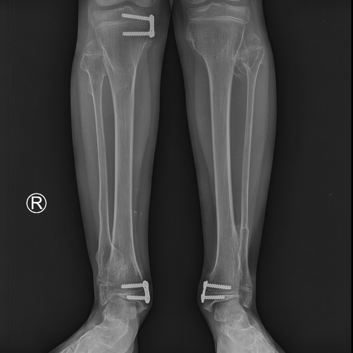
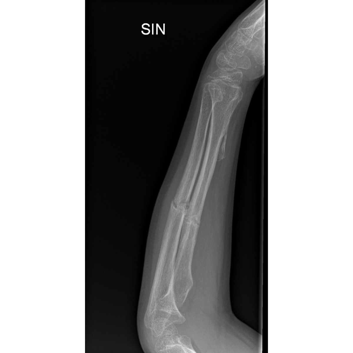
Multiple Hereditary Exostoses (MHE – multiple hereditary exostoses), also known as multiple osteochondromas, is a rare disease in which benign cartilage tumors that also produce bone tissue appear on long bones—most commonly in the femur and tibia.
MHE occurs in about 1 in 50,000 people, more commonly in men, and the growths usually develop during the first 10 years of life and continue to grow until puberty.
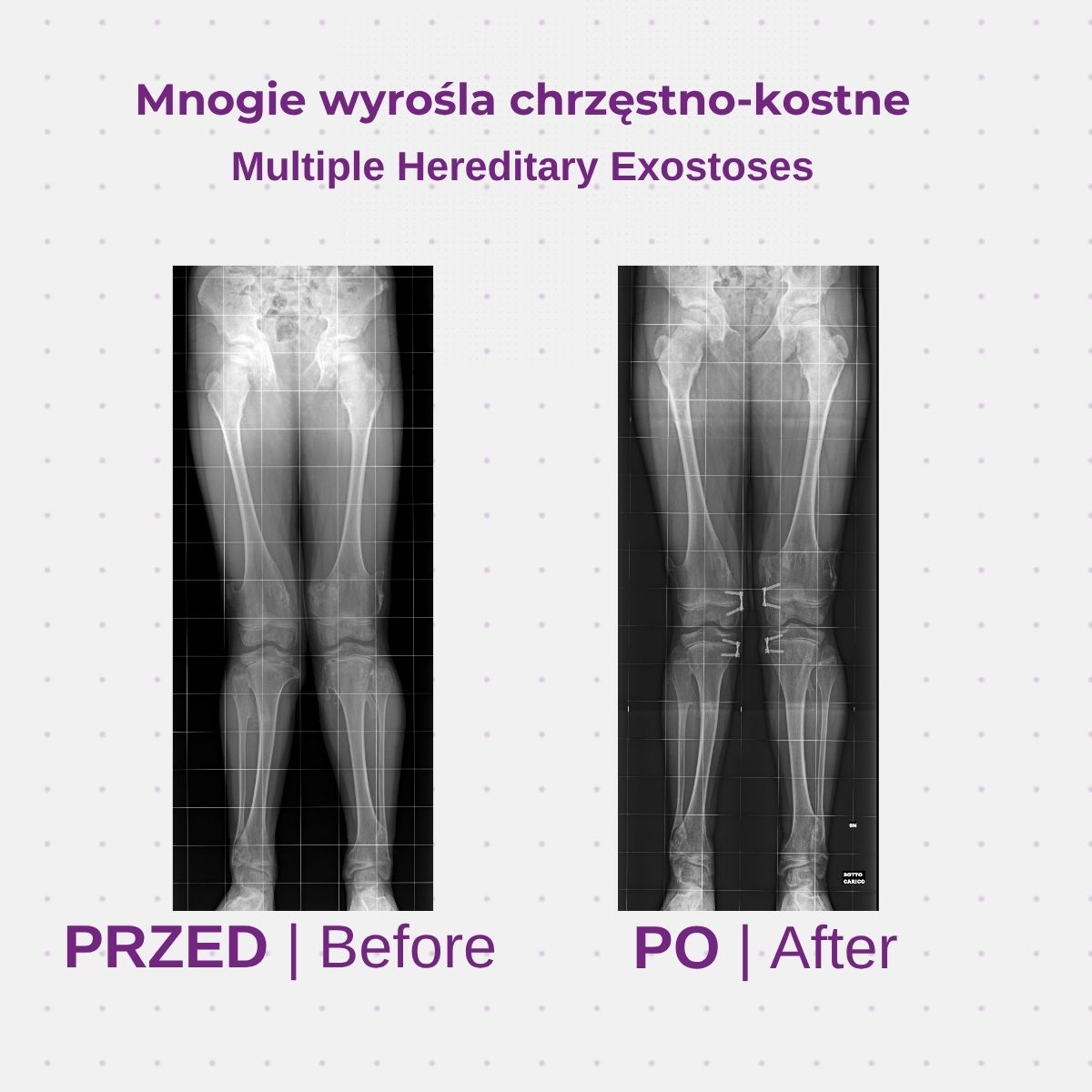
Multiple osteochondral growths?
Reasons
MHE is an autosomal dominant disorder; a single copy of the mutated gene is sufficient to cause the condition. Most often, the gene is inherited from the parents, although spontaneous mutations also occur. The disorder is associated with mutations in the EXT1 and EXT2 genes (and, according to the literature, also EXT3)—tumor suppressor genes responsible for the proper development and maintenance of bone and cartilage tissue.
Symptoms
Painless lumps near the joints, usually first noticed by parents
Discomfort or pain during physical activity, including nighttime pain that interferes with sleep
Numbness, tingling, and sensory disturbances resulting from pressure on the nerves
Changes in blood flow due to vascular compression
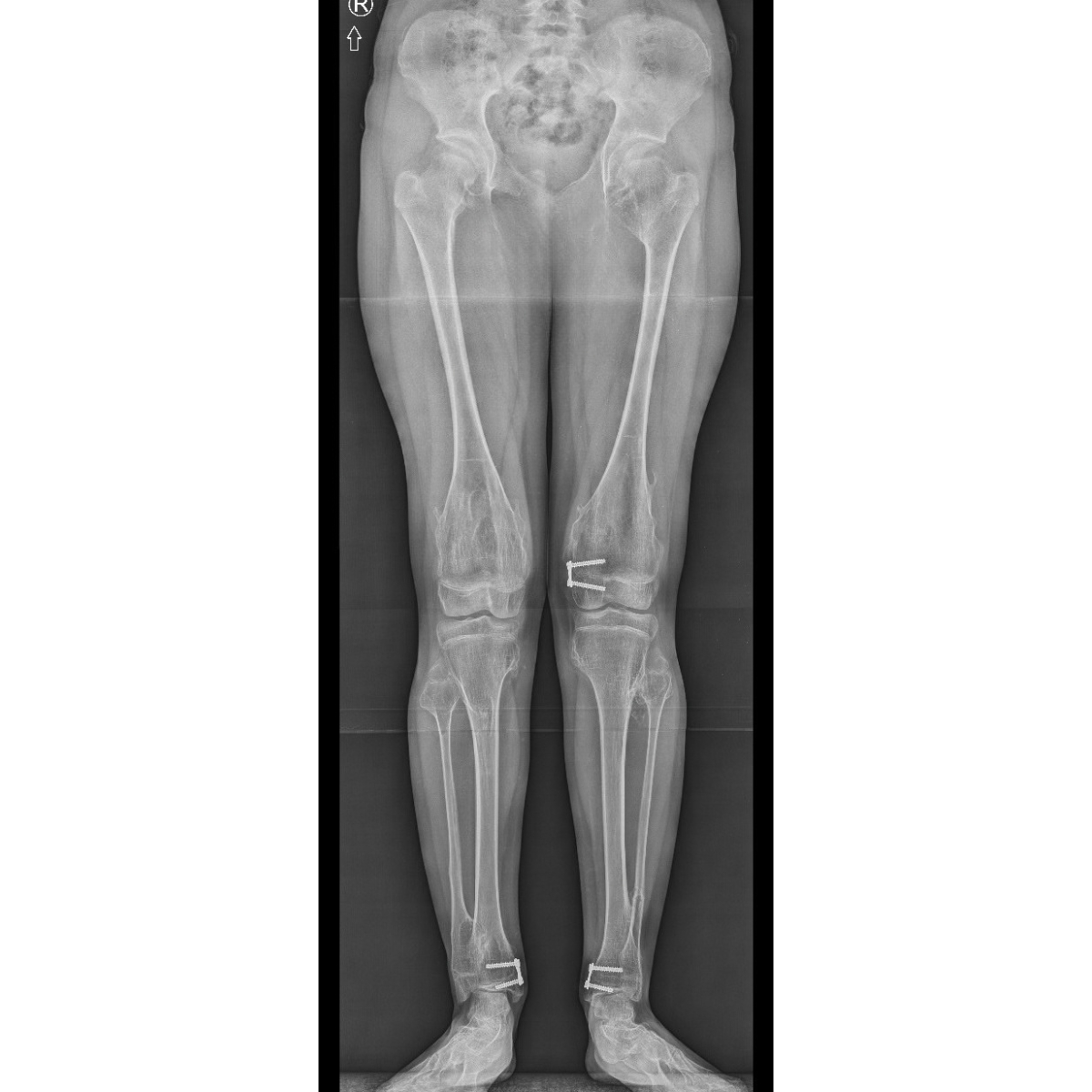
shortening and osteoarticular deformities that limit range of motion
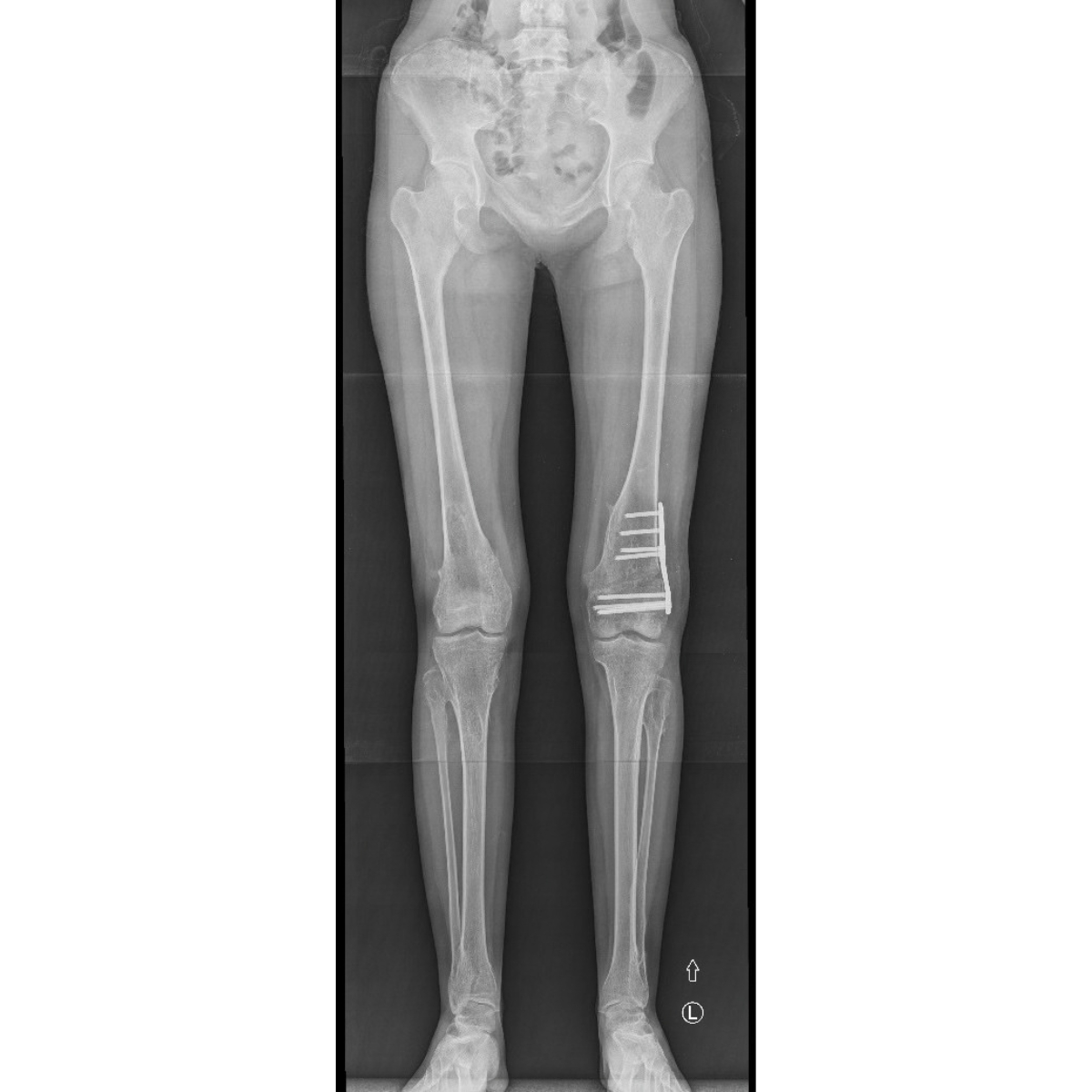
Limb length discrepancy (in 10–50% of patients, the discrepancy is 2 cm or more)
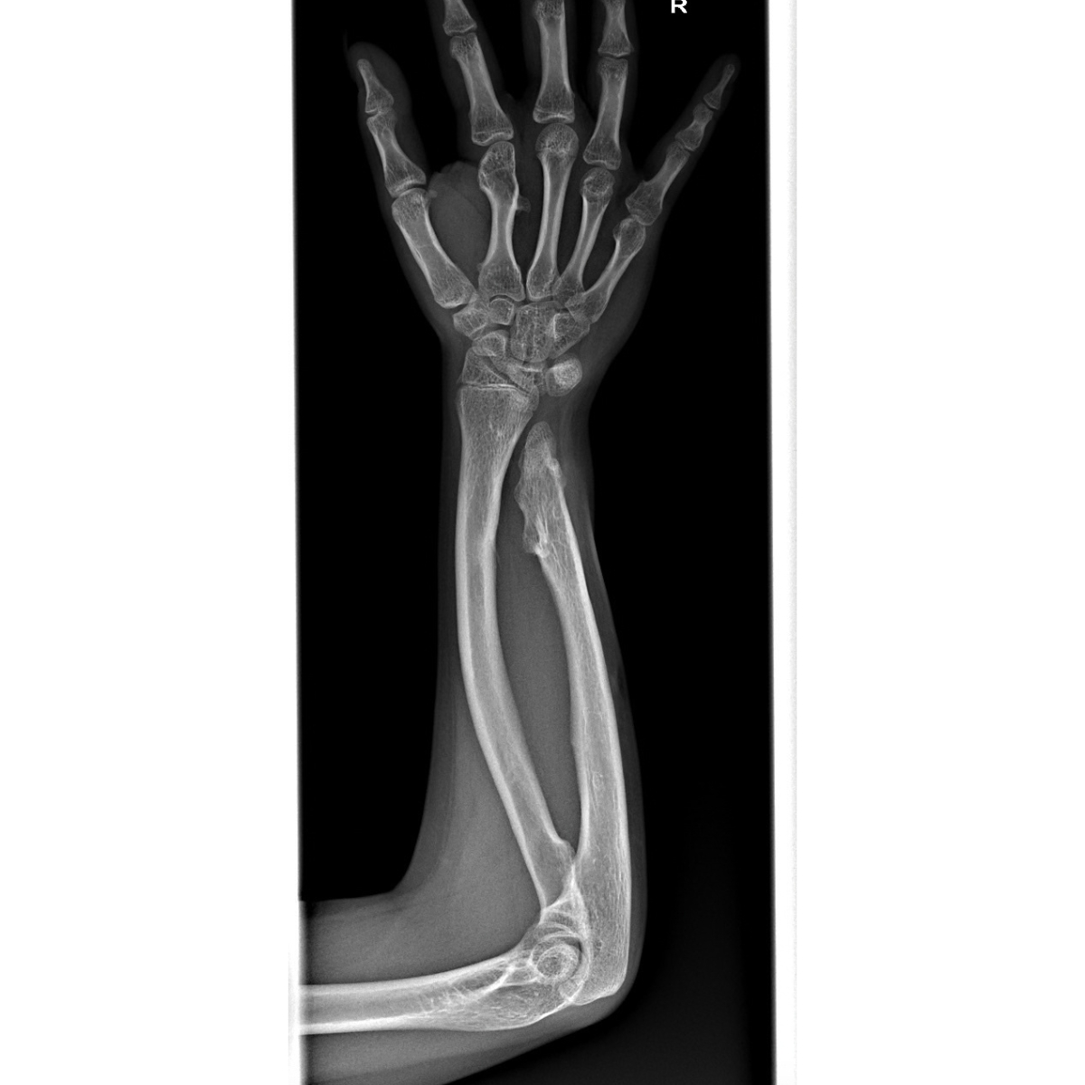
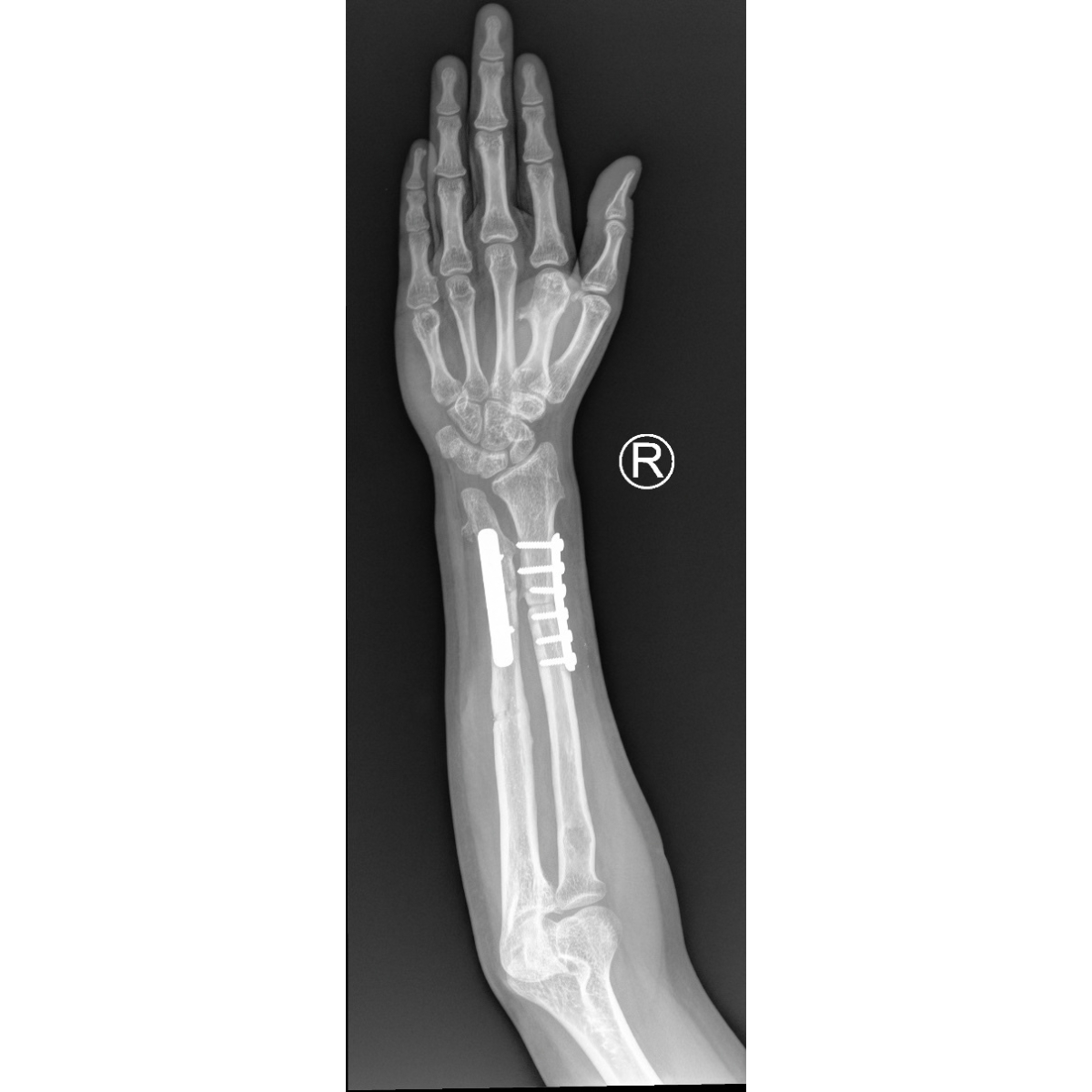
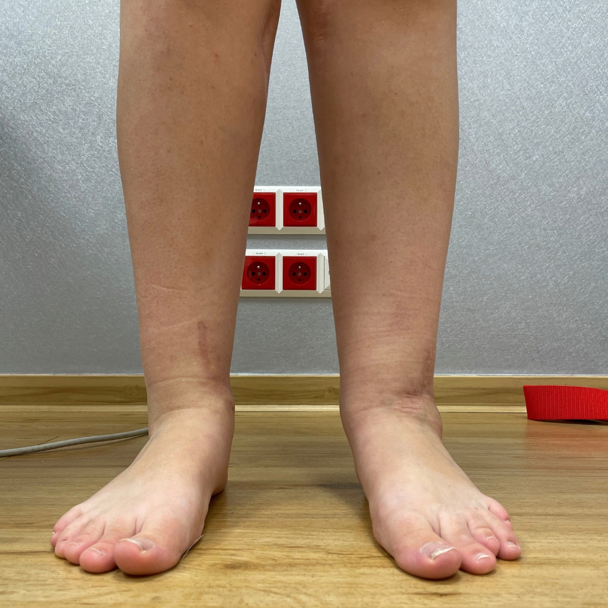
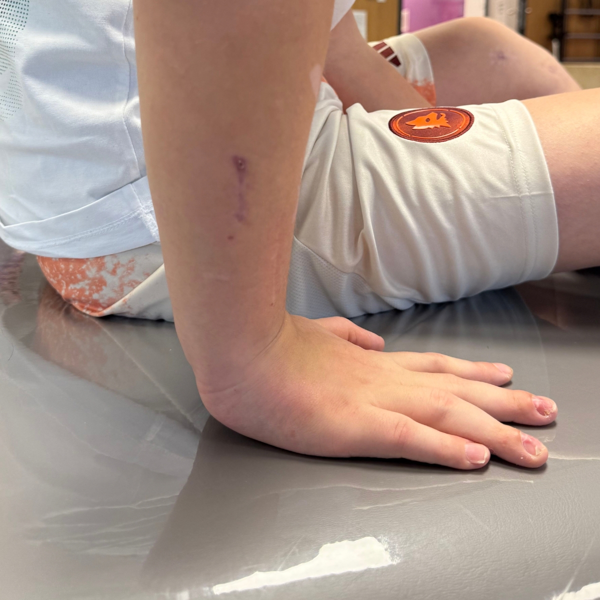
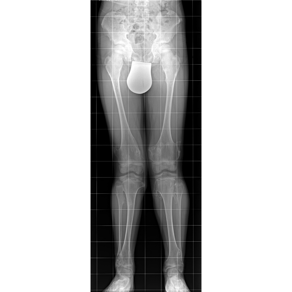
Bowlegs or knock-knees, forearm deformities
Diagnostics
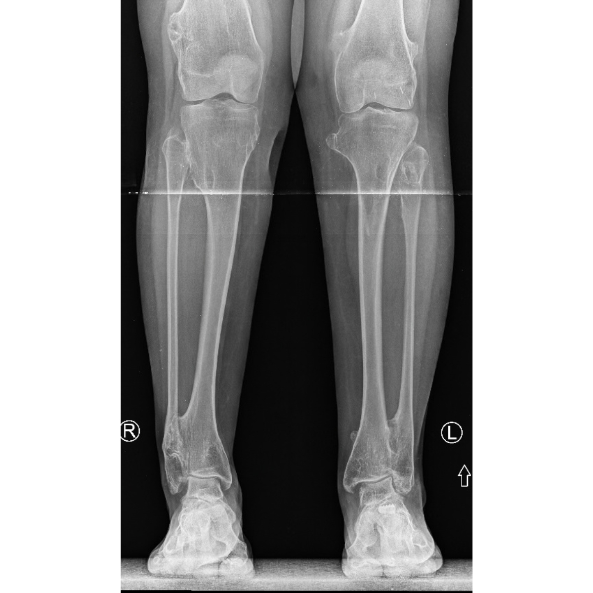
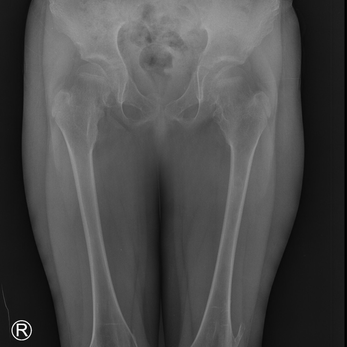
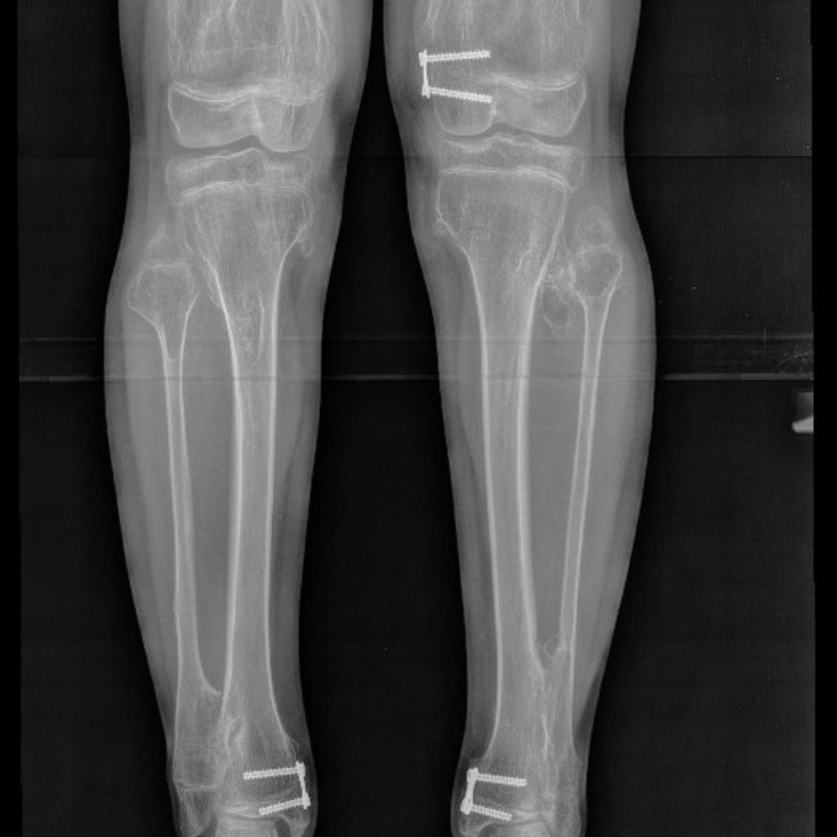
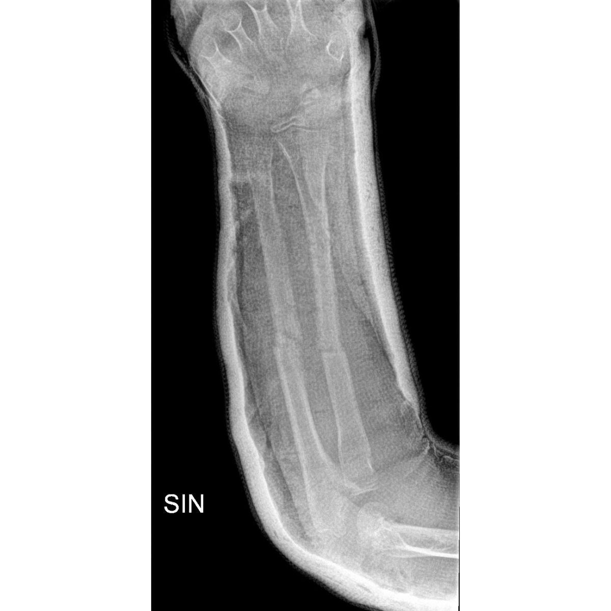
MHE is diagnosed based on a clinical examination, radiological imaging (X-rays, and in the case of larger, rapidly growing lesions, magnetic resonance imaging to assess the cartilaginous “cap” of the exostosis), and, if necessary, histopathological evaluation of a tumor biopsy specimen.
The global standard in the treatment of multiple osteochondral lesions
Paley European Institute Support
Our team specializes in the treatment of rare skeletal disorders in children and adults, combining advanced diagnostic imaging with a personalized surgical and rehabilitation treatment strategy.
Stages of Treatment
From the initial consultation and monitoring of changes over time (regular X-ray follow-ups), through qualification for surgery in cases of pain, rapid growth of bone spurs, or risk of malignancy, to the correction of limb axial deformities and length equalization.
Comprehensive Care
Treatment includes not only the removal of bone spurs, but also the correction of deformities, limb lengthening, and comprehensive postoperative physical therapy—all under the care of a single team of specialists.
When should you see a specialist?
when a child develops painless lumps near the joints, which are noticeable during a physical examination or through everyday observation
when pain occurs during physical activity or when nighttime pain disrupts sleep
when there is numbness, tingling, or other sensory disturbances in the limbs
when there is a visible difference in leg length or a deformity of the knee axes (knock-knees/bowlegs)
When a growth begins to grow rapidly or is accompanied by increasing pain—in such a situation, a consultation and possible surgery should take place immediately due to the small but real risk of malignancy (approximately 1–2% of cases)

How do we treat multiple osteochondral lesions?
Treatment for MHE is tailored to each individual and may include observation, conservative management of symptoms, or surgical intervention.
Diagnostics and Planning
A clinical and radiological study evaluating the number, location, and size of growths
Assessment of limb alignment and length, and the risk of neurological complications
Planning a treatment strategy tailored to the patient's age and stage of growth
Post-operative care
Surgical removal of growths that compress nerves or blood vessels or restrict movement
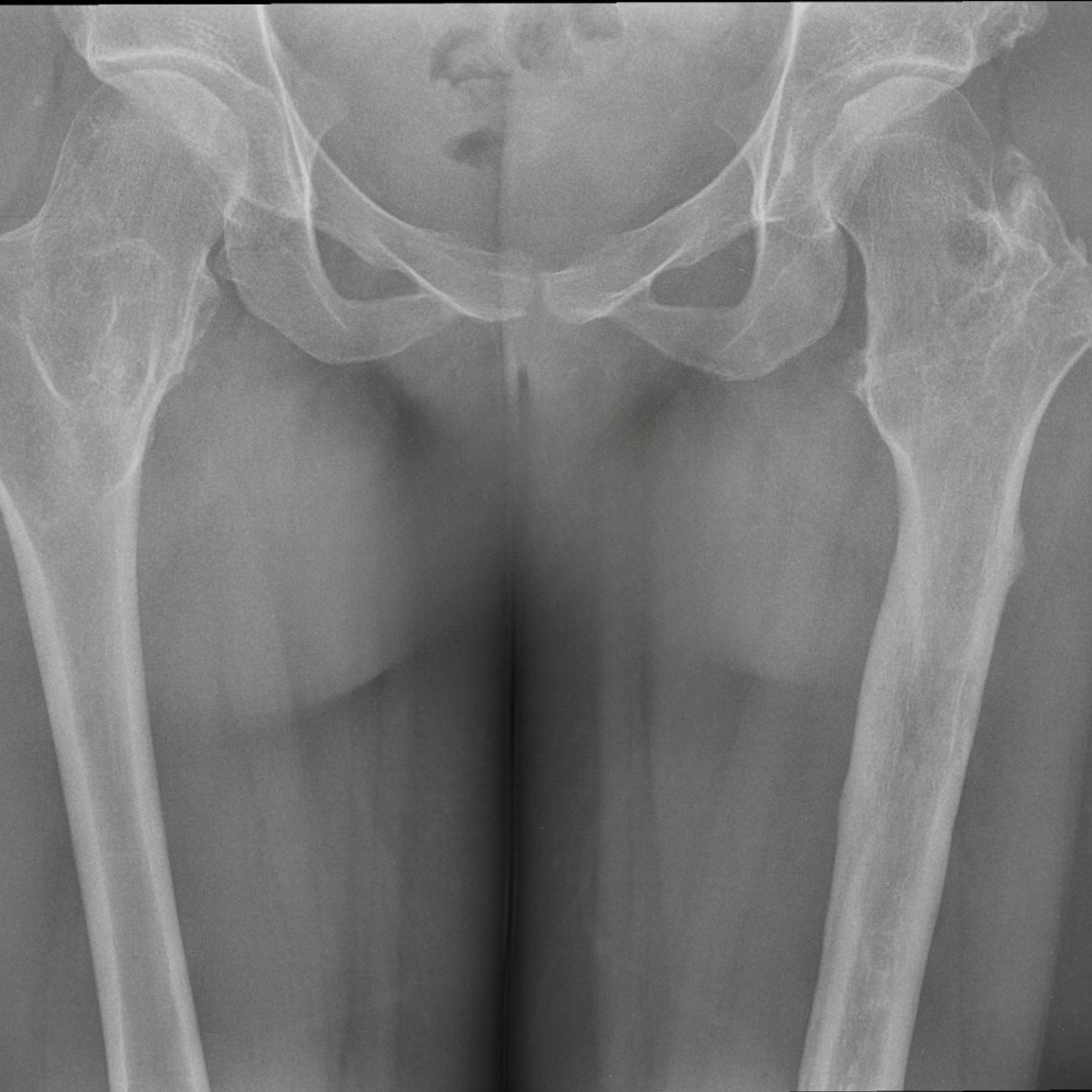
Femoral osteotomy or removal of osteophytes from the femoral neck in cases of hip lesions
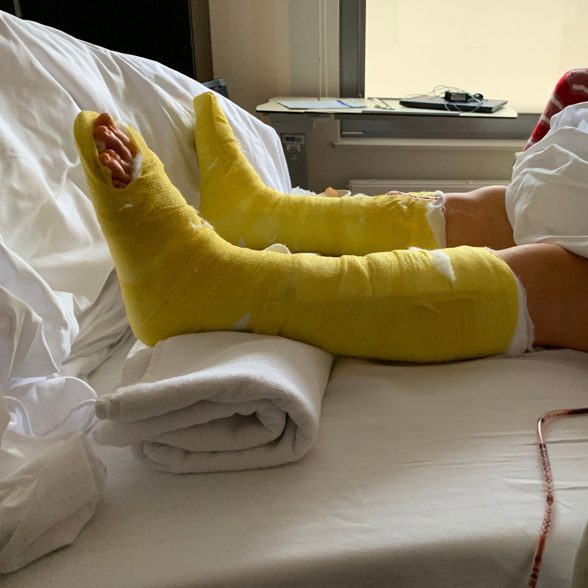
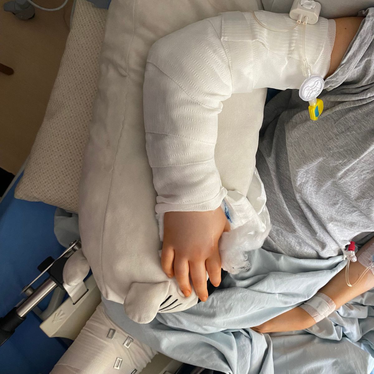
A recovery period lasting approximately 2–3 weeks, during which one should avoid straining the area that underwent surgery
Limb lengthening
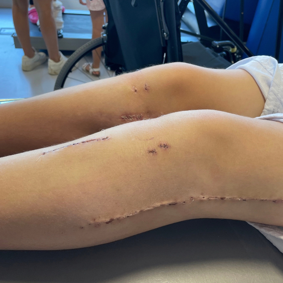
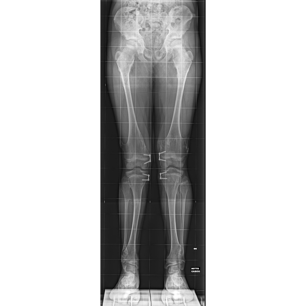
Hemiepiphysiodesis—partial, controlled closure of the growth plate to correct the alignment of the knee
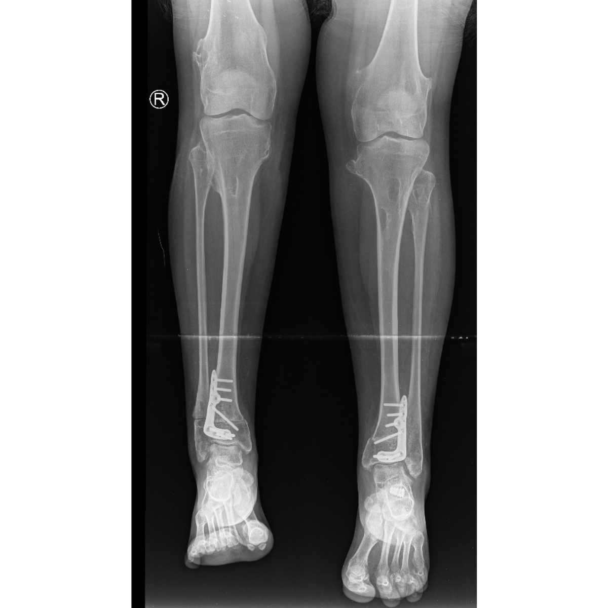
In cases of severe axial malalignment and in adults—bone lengthening and angle correction using an external fixator
Rehabilitation and Long-Term Care
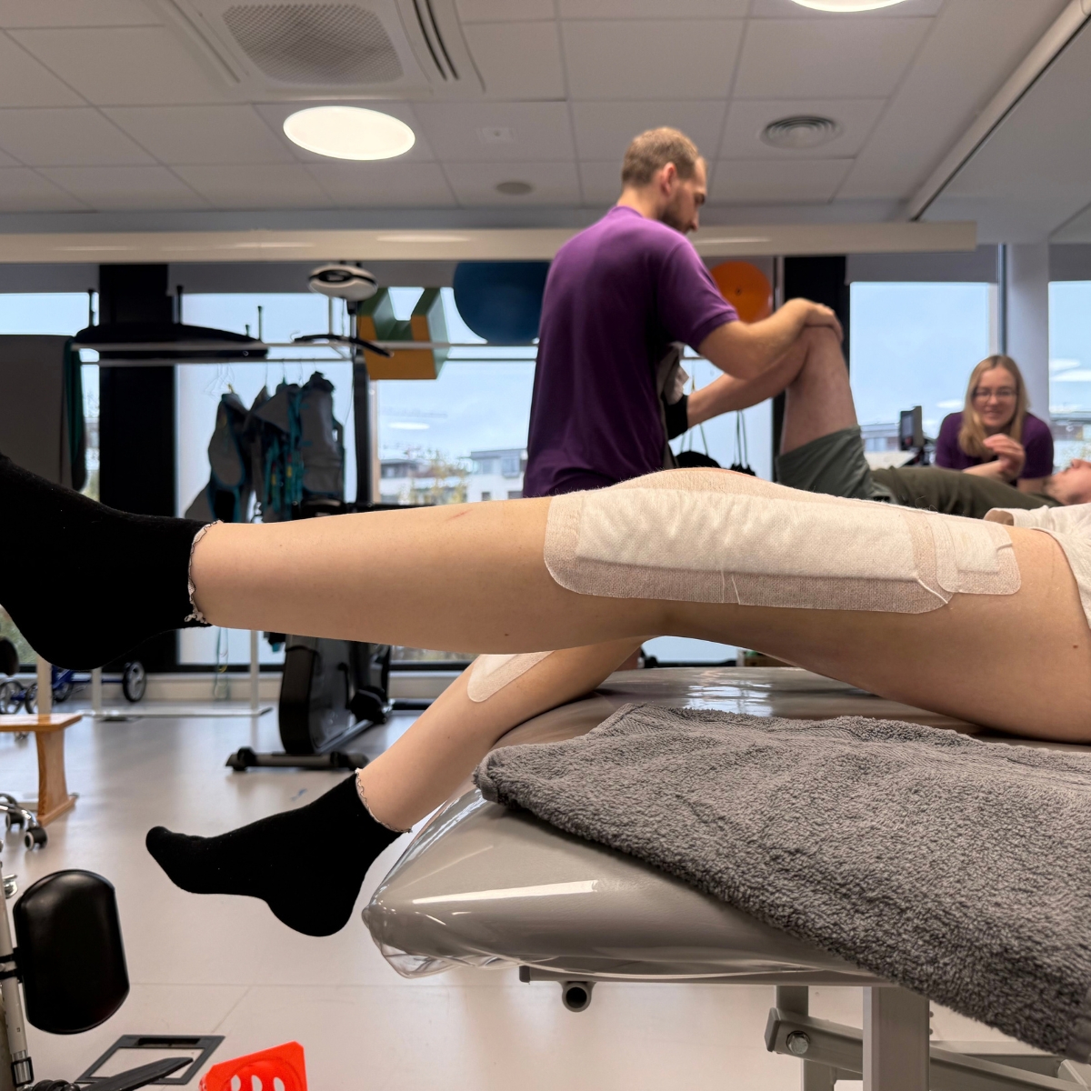
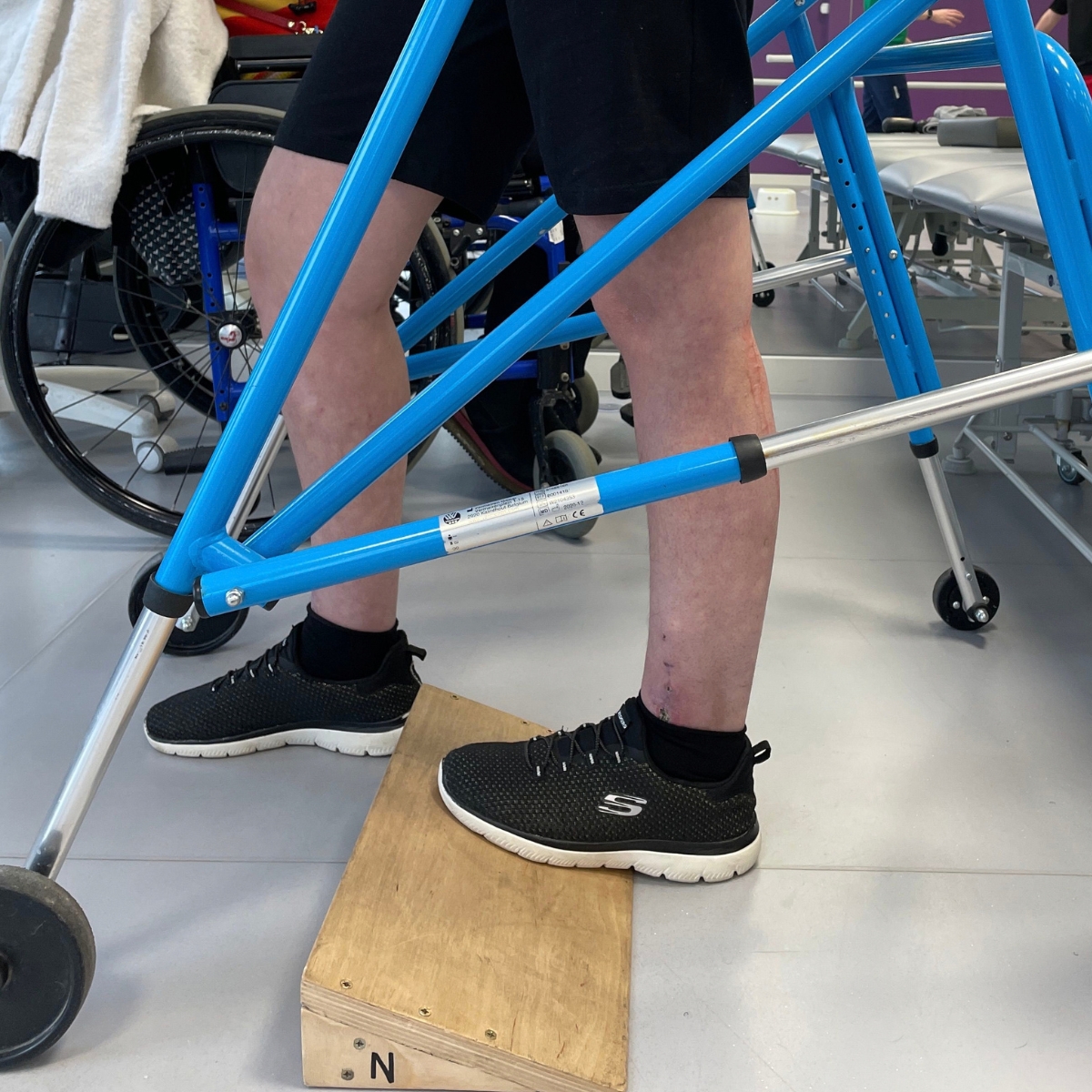
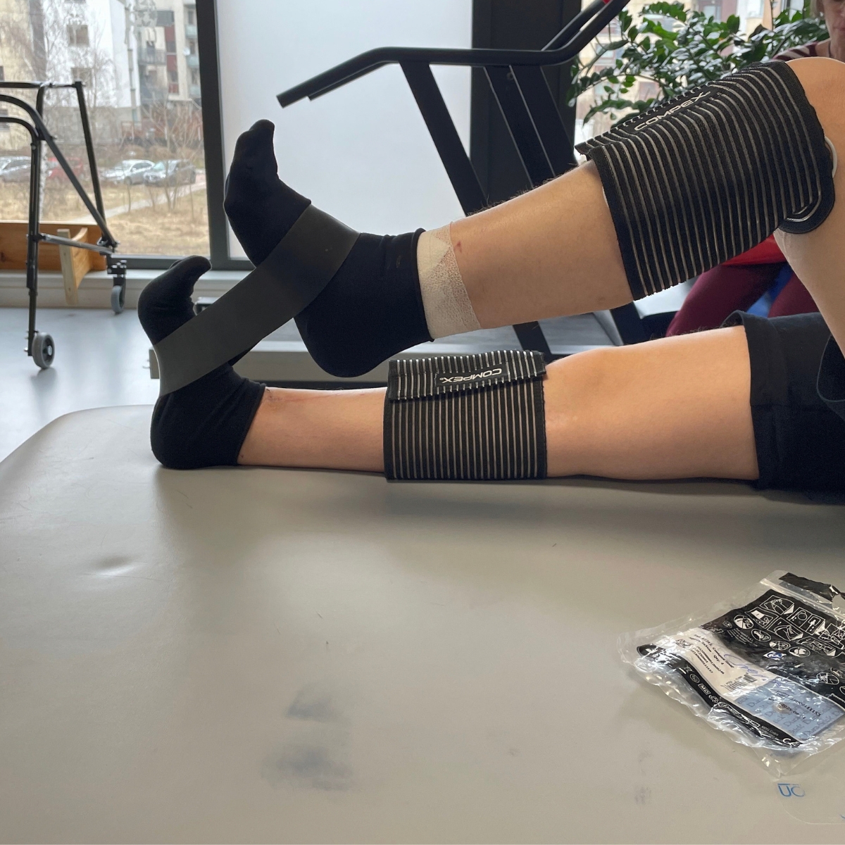
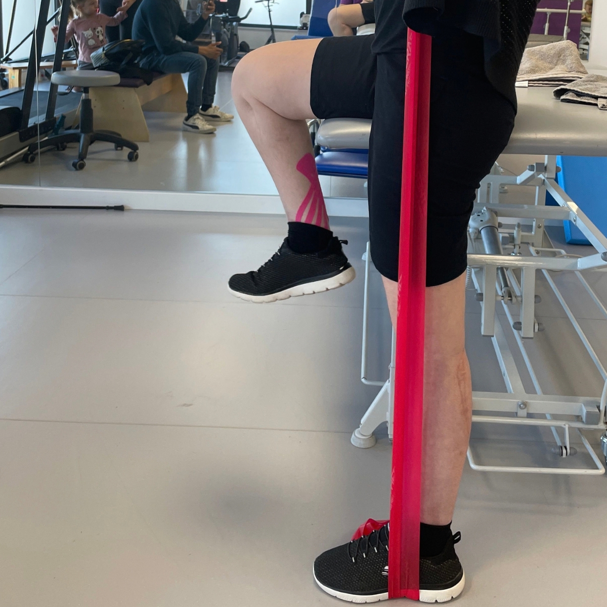
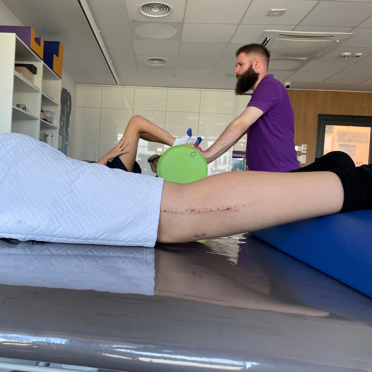
Physical therapy to support a return to full fitness after surgery
Regular imaging examinations to monitor for any new changes
Supporting patients and their families in their daily lives with the disease
What challenges do patients with MHE face?
Limited mobility —growths can change the range of possible movements from one day to the next; activities such as walking, running, bending, carrying, or grasping may become difficult.
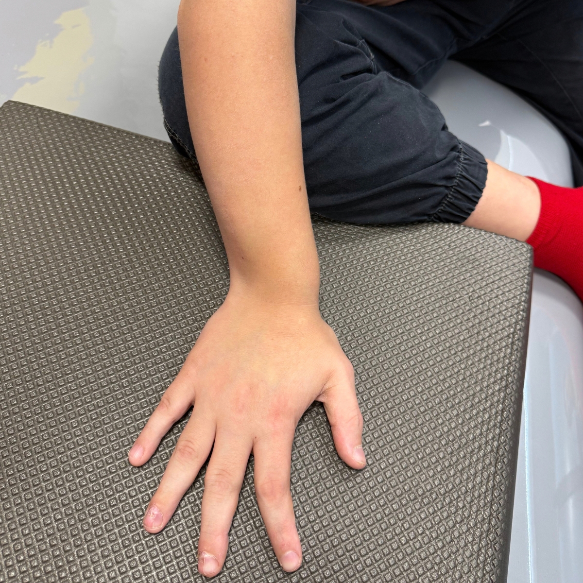
Manual difficulties – patients with hand and arm involvement may have trouble tying their shoes, writing for long periods, throwing, or swimming certain strokes.
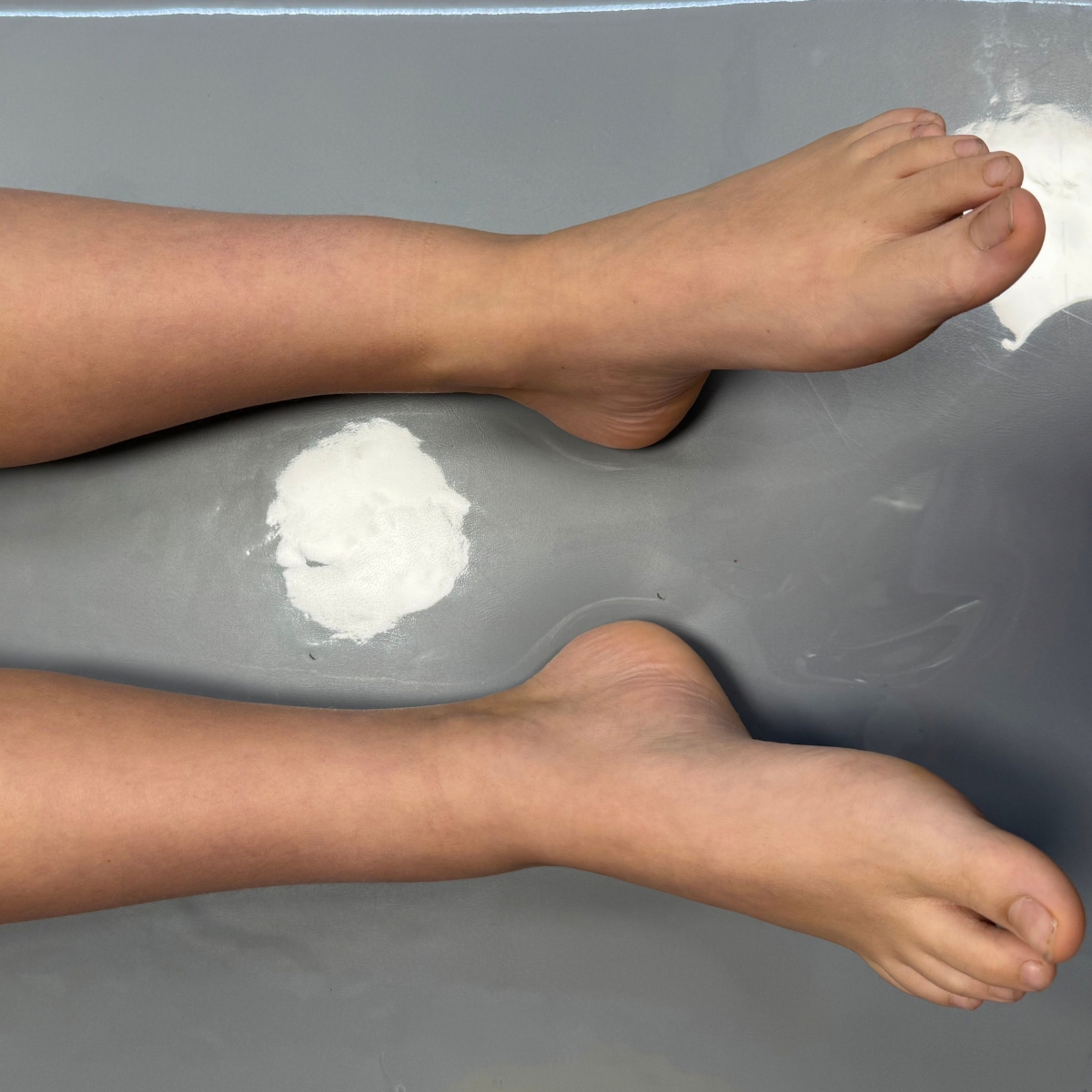
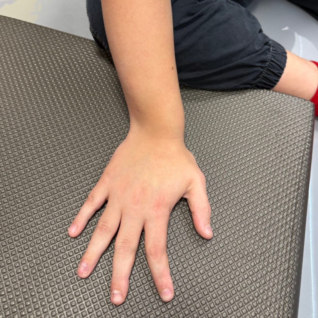
Foot deformities – People with lower-limb involvement often have deformed feet with toes of varying lengths, which causes pain and gait abnormalities.
Fatigue – Limited mobility and pain can lead to getting tired more quickly and the need for longer periods of rest.
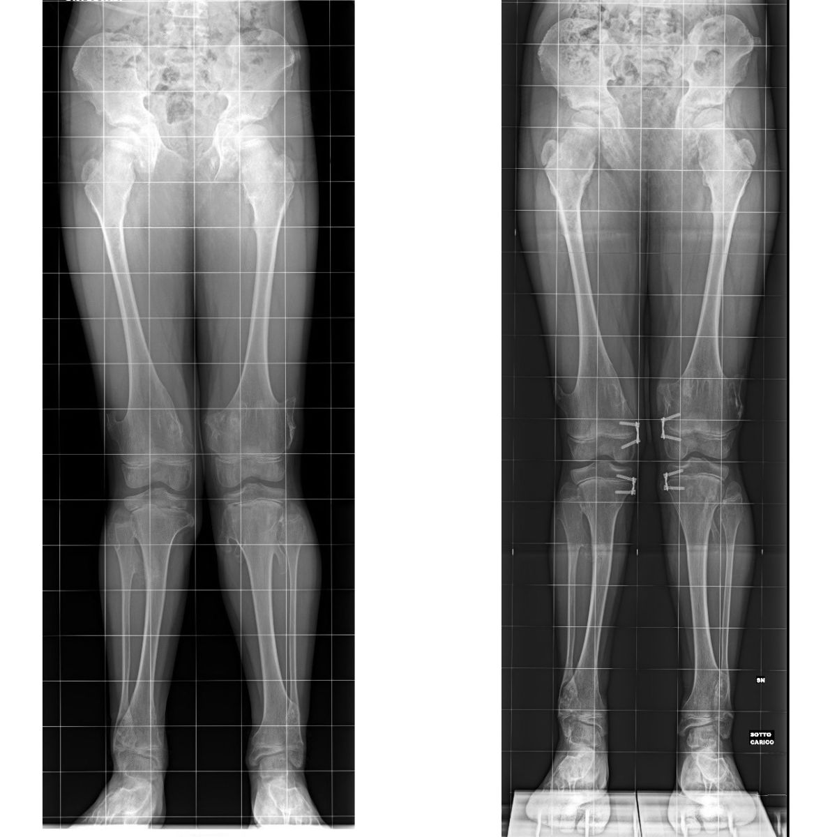
Bony Exostoses in the Femur
The femur is affected by osteochondral lesions twice as often as the tibia, making it one of the most important areas to monitor in patients with MHE.
Appearance and location —growths can appear in the area of both the knee and hip joints, including the femoral neck.
Axial deformities —involvement of the femur is often associated with the development of genu varum or genu valgum and limb length discrepancy.
Conservative treatment —regular radiological monitoring of lesions that do not cause symptoms.
Surgical treatment —depending on the location and severity of the lesions, femoral osteotomy, removal of femoral neck exostoses, or hemiepiphysiodesis to correct the knee axis may be performed.
Limb lengthening —in cases of significant leg length discrepancy, external devices are used to enable controlled and precise bone lengthening.
You should entrust your health and that of your children to professionals who know exactly how to manage rare diseases. Please contact us to schedule a consultation.
Our Experts Meet the Paley European Institute Team

Family-Centered Care: treatment in which the family is part of the care team
Family-Centered Care(FCC) is the treatment model used at the Paley European Institute. In this model, the family of a child with a congenital limb defect is not merely an observer but a full-fledged member of the treatment team. Treatment for congenital limb defects often lasts for months or years and involves several surgeries, limb lengthening, and months of rehabilitation. Therefore, parents who understand the treatment plan and feel that their concerns are being heard have a real impact on its outcomes.
What does family-centered care look like in practice?
Parents do not wait in the hallway—while their child is undergoing surgery, the Hospital Services Coordinator and a physician’s assistant keep parents updated on the progress of the procedure via WhatsApp. During this time, a psychologist stays by the parent’s side to support them through this most difficult hour.
One treatment plan instead of multiple visits—during the consultation, specialists from various fields meet with the child in one place and develop a comprehensive treatment plan. The conversation begins not with the question “What hurts?”, but with what the child likes to do and what the family’s daily life is like.
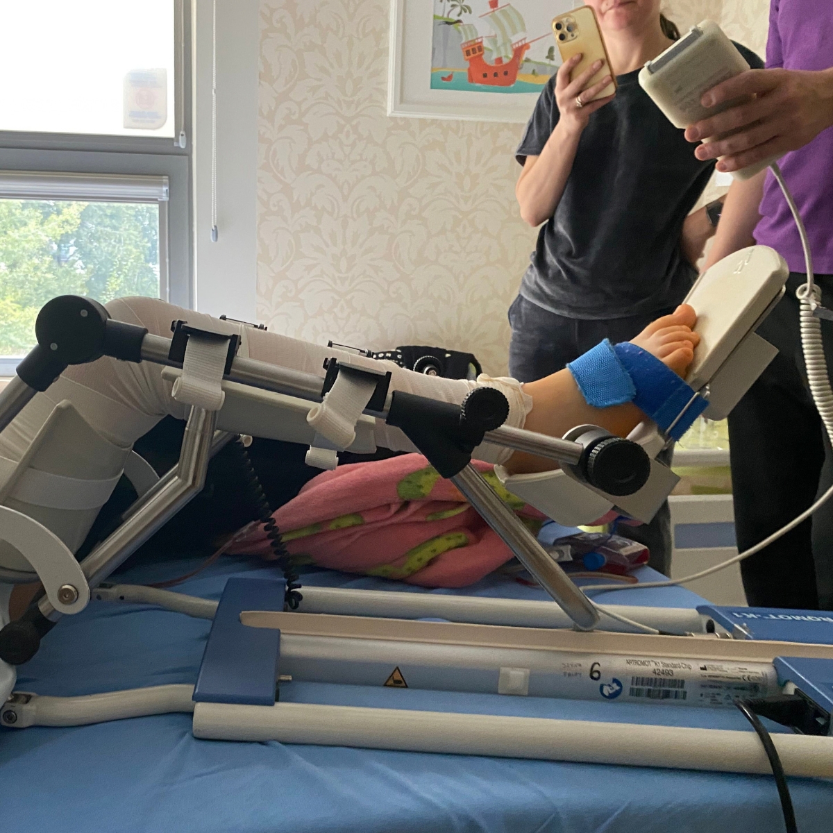
The same physical therapist before, during, and after surgery—the therapist present in the operating room—conducts rehabilitation starting on the first day of recovery. The child exercises with someone they already know, which reduces anxiety and facilitates a return to physical fitness. This is particularly important during limb lengthening, when systematic physical therapy is crucial for maintaining joint mobility.
Comprehensive care in one place—X-rays, blood tests, an orthotist, a psychologist, a dietitian, a speech therapist, and certificates for schools and government offices—are all available without unnecessary referrals or having to search for other facilities. Surgeries are performed at our partner hospital, Medicover, where the Paley European Institute team cares for the patient from admission through discharge.
Support for families from other cities and countries—families traveling here for medical treatment receive assistance with arranging transportation, accommodations, and all the logistics of their stay.
Parents' Council—parents who have themselves undergone treatment at the Paley European Institute—regularly test and evaluate new approaches, helping to shape the model of care.
Why does family involvement improve treatment outcomes?
Parents who understand the treatment plan are better able to follow rehabilitation recommendations at home and recognize signs that require contacting a doctor more quickly. Thanks to preparing the family for home care, children who undergo surgery at the Paley European Institute stay in the hospital for an average of only 2 days. A shorter hospital stay and less stress lead to a more peaceful recovery and a better quality of life for the entire family.

Multimedia
Related articles
Read the blog posts on this topic

Rare Diseases Day - MHE

The Paley European Institute has partnered with the Multiple Chondrosarcoma (MHE) Association to help patients with the syndrome.

Multiple cartilaginous outgrowth syndrome MHE
Congenital hereditary cartilaginous outgrowths

Everything you need to know about the causes, symptoms and treatment of multiple cartilaginous outgrowths!
Your most frequently asked questions
In Other Words: What You Should Know Before Deciding on Treatment.
MHE (multiple hereditary exostoses) is a rare, hereditary disorder in which numerous benign tumors composed of cartilage and bone tissue develop on the long bones—most commonly on the femur and tibia. On average, each patient has between 15 and 18 exostoses.
Yes. MHE is inherited in an autosomal dominant pattern—a single copy of the mutated gene (most commonly EXT1 or EXT2) is sufficient for the disease to manifest. The gene is usually inherited from one of the parents, although spontaneous mutations also occur.
The disease is very rare—it affects about 1 in 50,000 people, most often men.
The most common initial sign is painless, firm lumps around the joints, which can be felt in the child’s hands or legs. These may be accompanied by pain during physical activity or at night, numbness and tingling, limited joint mobility, as well as a visible difference in limb length or knee misalignment.
In a very small percentage of cases (about 1–2%), the tumor may become malignant and develop into a chondrosarcoma. That is why regular radiological follow-up and orthopedic consultations are so important—they allow for the early detection of concerning changes.
The diagnosis is based on clinical and radiological (X-ray) examination and, if necessary, magnetic resonance imaging (MRI), which allows for the evaluation of the cartilaginous “cap” of the exostoses that is not visible on an X-ray. In certain cases, a histopathological evaluation of a tumor biopsy specimen is necessary.
No. Most growths are asymptomatic and require only regular follow-up over time. Surgery is considered when the growth causes pain, puts pressure on nerves or blood vessels, limits mobility, leads to limb deformity, or grows at an unusually rapid rate.
Meet Our Patients