Lower Limb Deformities – Causes, Symptoms, and Treatment
Lower limb alignment abnormalities are one of the most common orthopedic problems in children, though they can also affect adolescents and adults. The Paley European Institute offers precise diagnostics and treatment based on methods developed by Dr. Dror Paley, a world leader in the correction of limb alignment disorders.
What are limb deformities?
Lower limb deformities encompass a wide spectrum of disorders, from deviations in the limb axis (valgus, varus), through leg length differences, to abnormal rotation of the femur and tibia. Some of these are a natural part of a child's development and resolve spontaneously, while others require early intervention to prevent joint overload and permanent degenerative changes.
An incorrect limb axis causes an uneven distribution of forces acting on the knee and hip joints, which over time leads to overloading of the joint cartilage, damage to the meniscus and accelerated development of degenerative disease.
Deformities of the Lower Limbs
Reasons
Overweight and Obesity
Rickets
Kidney Diseases
Asymmetric closure of growth plates following injuries
Inflammation
Blount's Disease
Growth Disorders in Childhood (in Adults)
Metabolic Bone Diseases
Severe osteoarthritis
Injuries or fractures
Symptoms
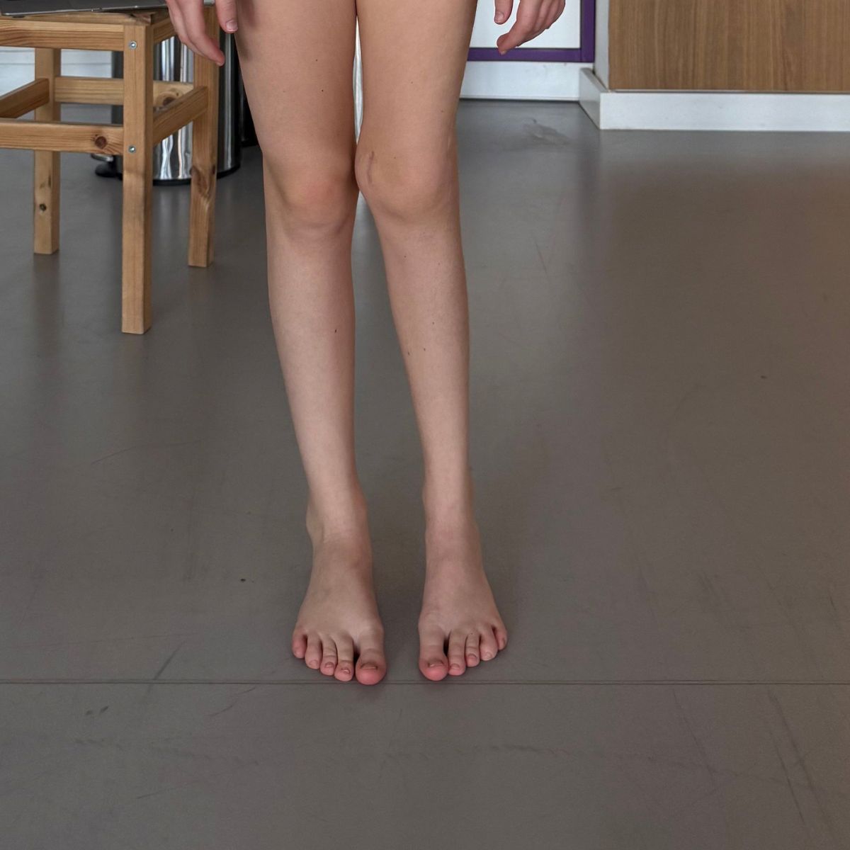
Visible valgus or varus deformity of the knees that exceeds age-appropriate norms
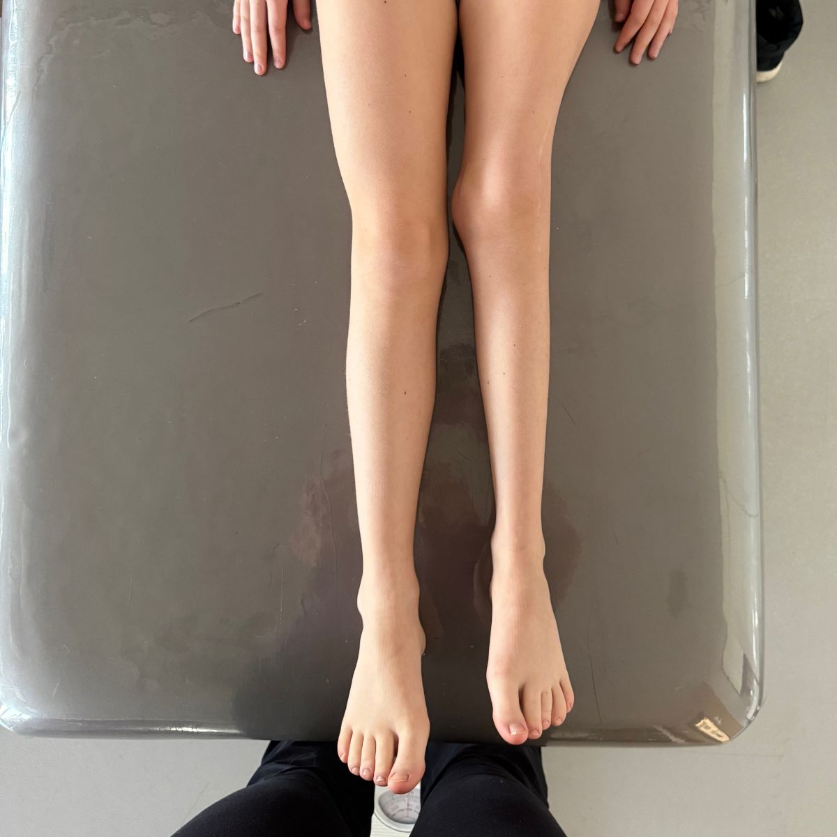
Uneven leg lengths
Abnormal, compensatory gait pattern
Knee, hip, or back pain caused by joint overuse
Progressive Degenerative Joint Changes in Adolescents and Adults
Asymmetry in leg alignment visible when standing
Diagnostics
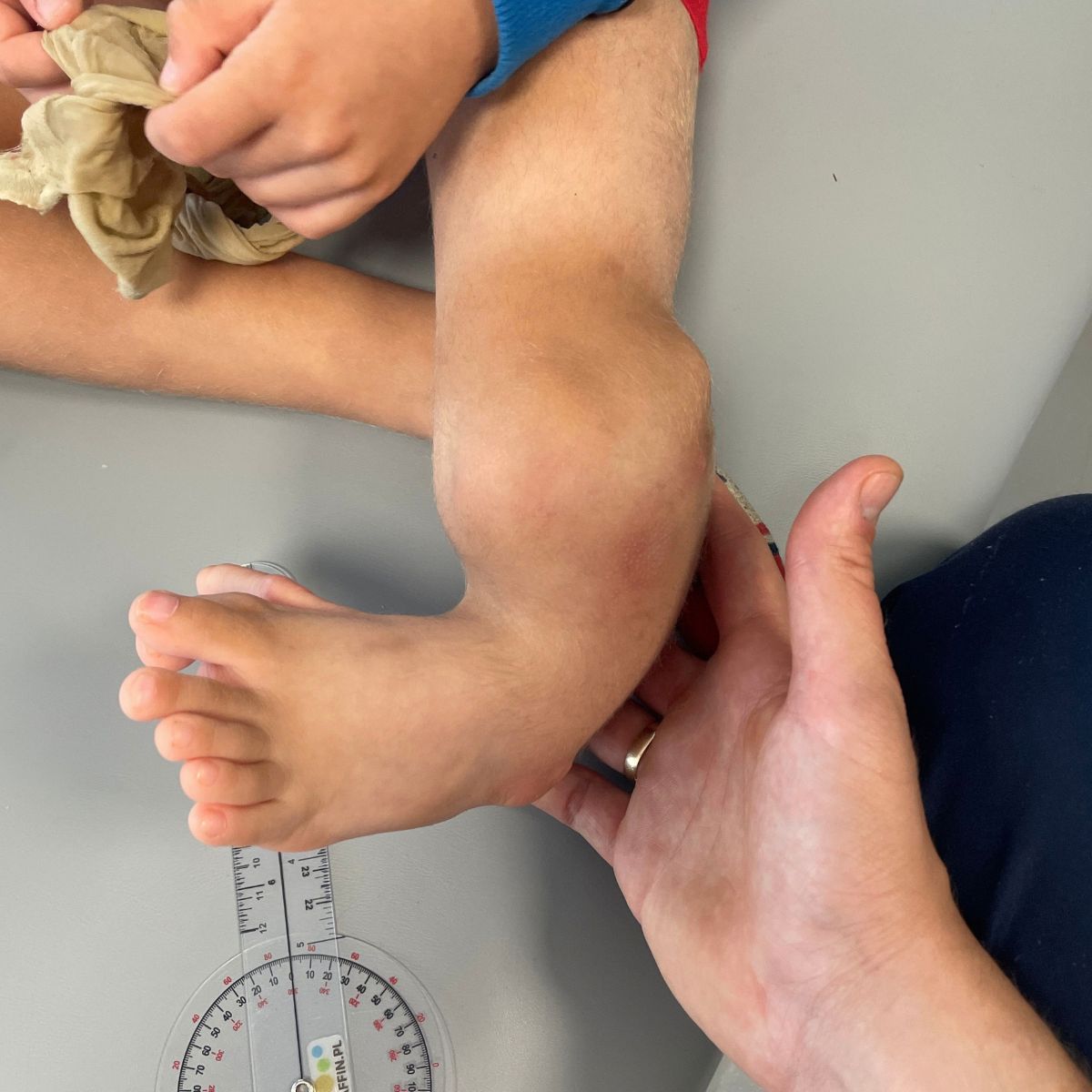
Detailed Clinical Examination and Assessment of Gait Patterns
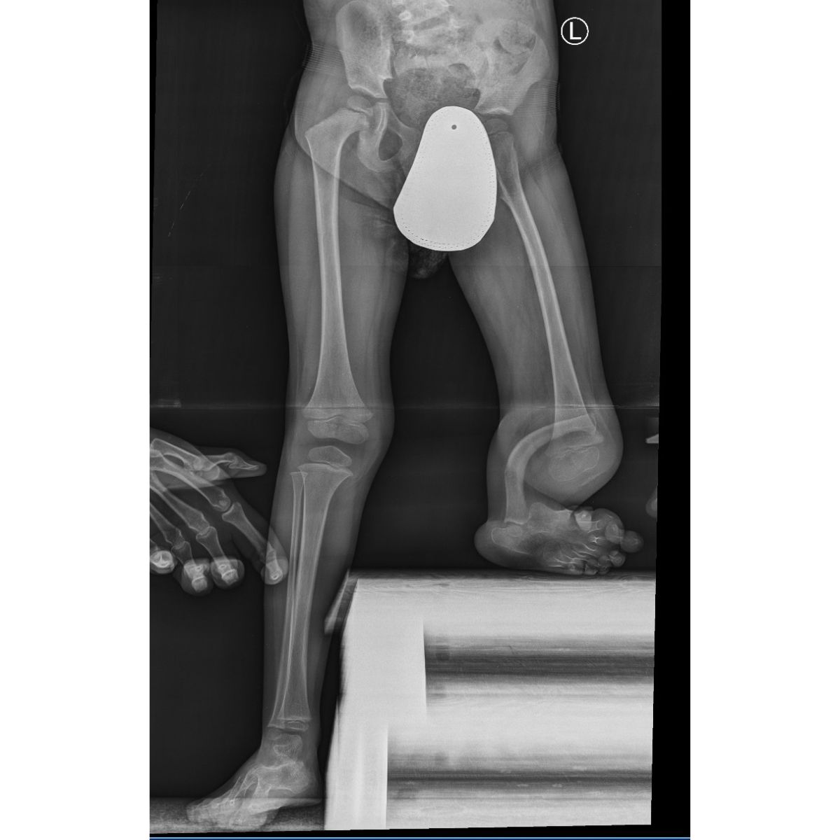
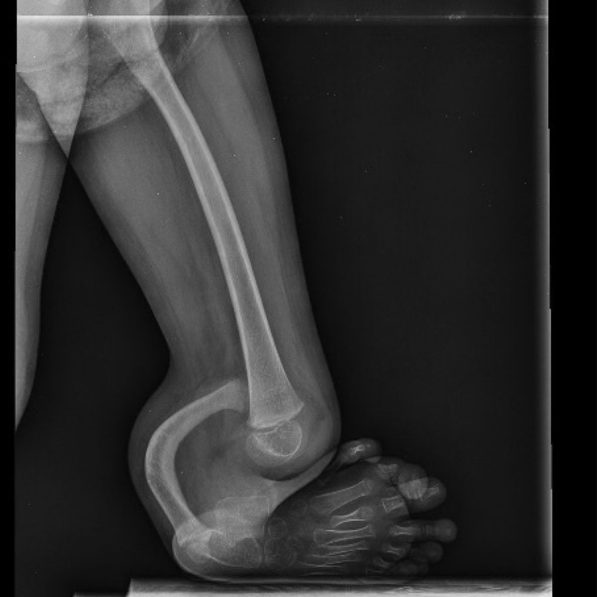
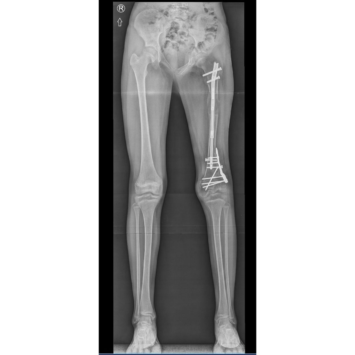
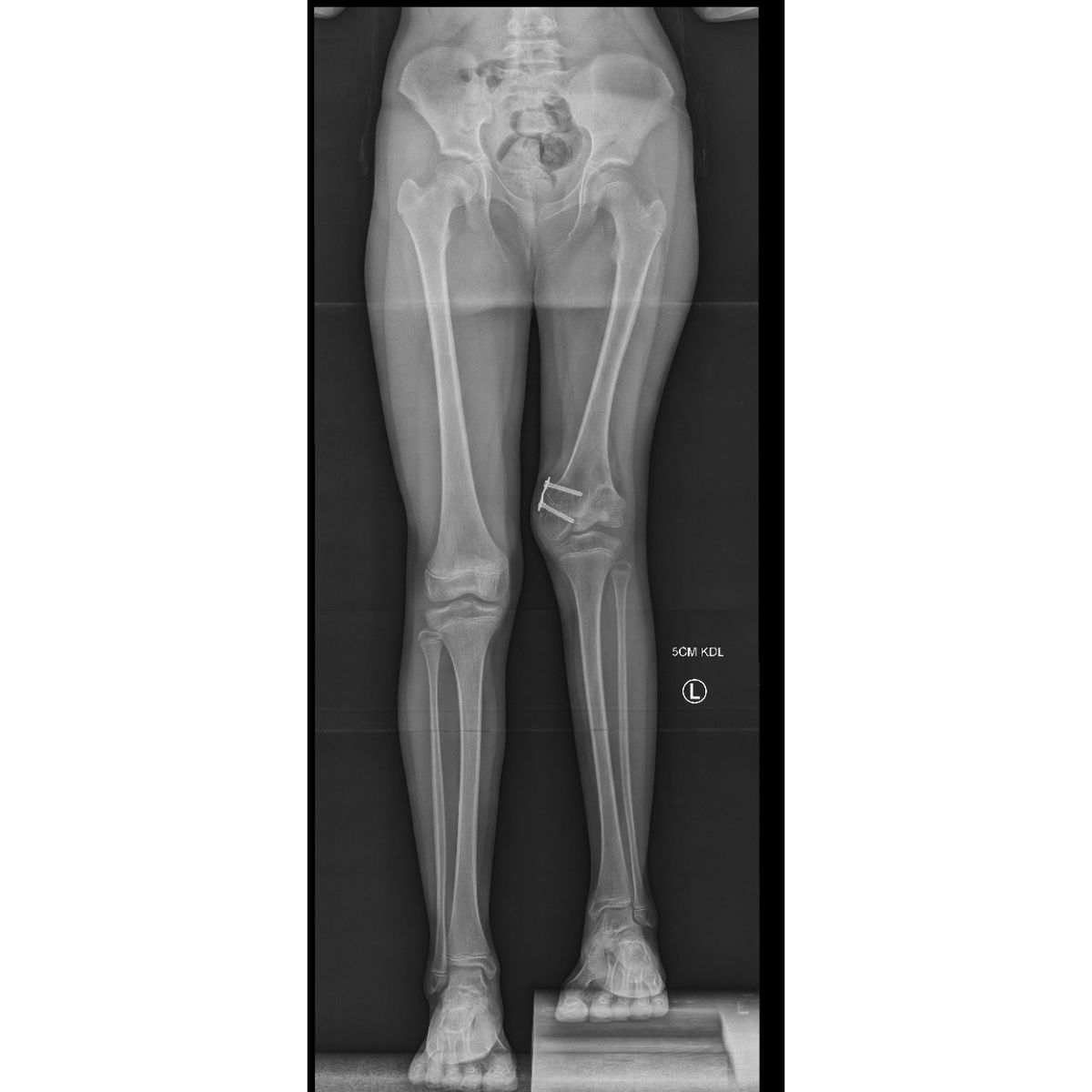
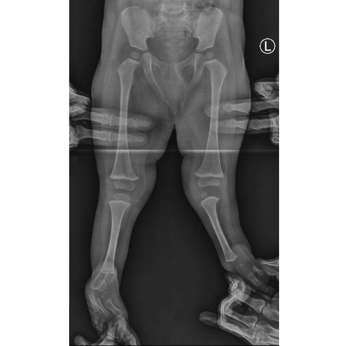
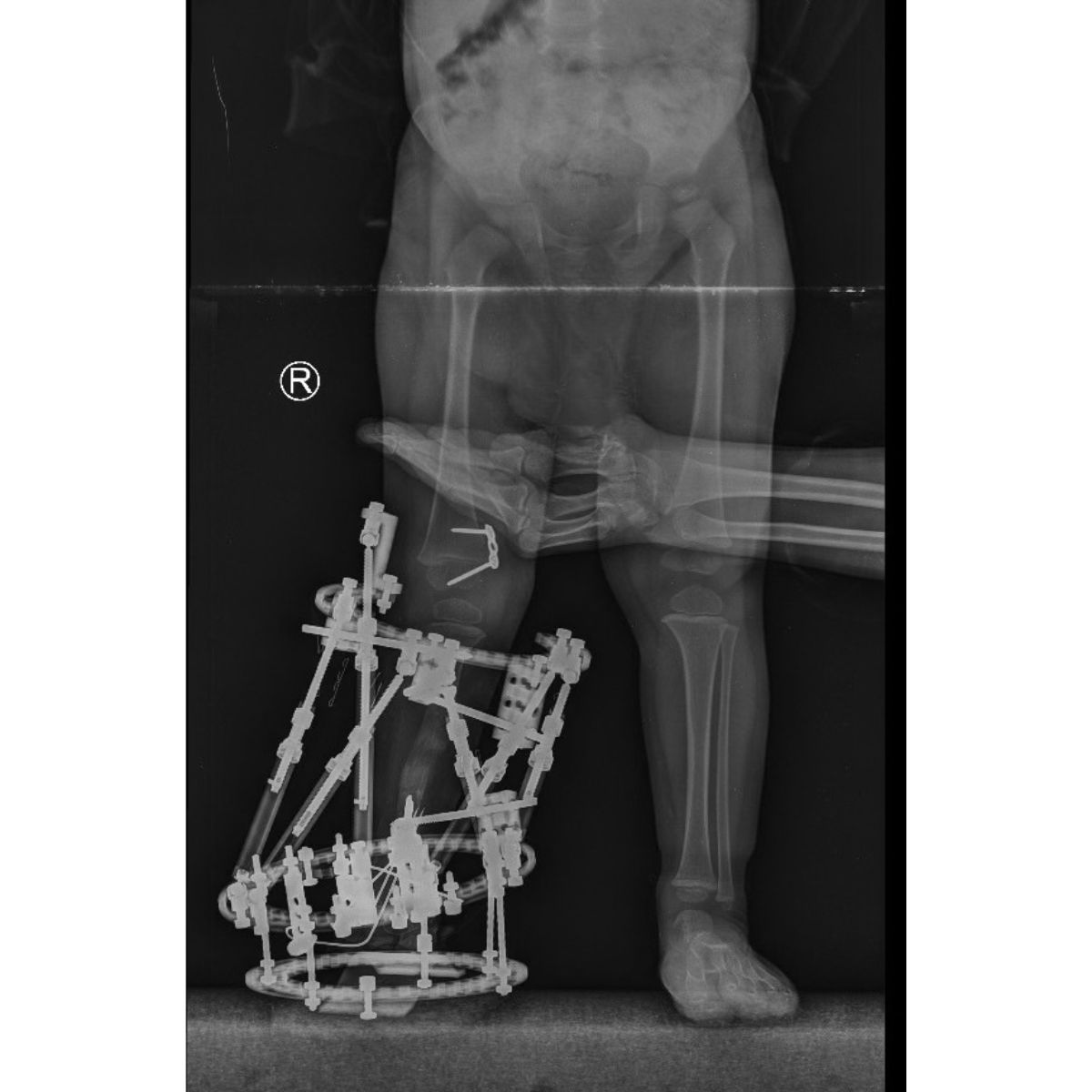
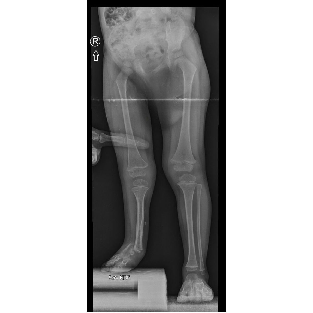
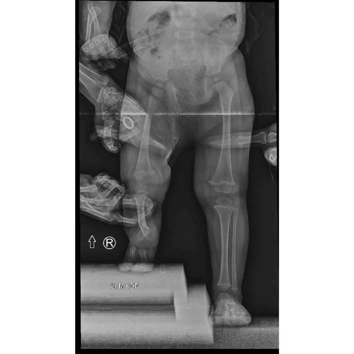
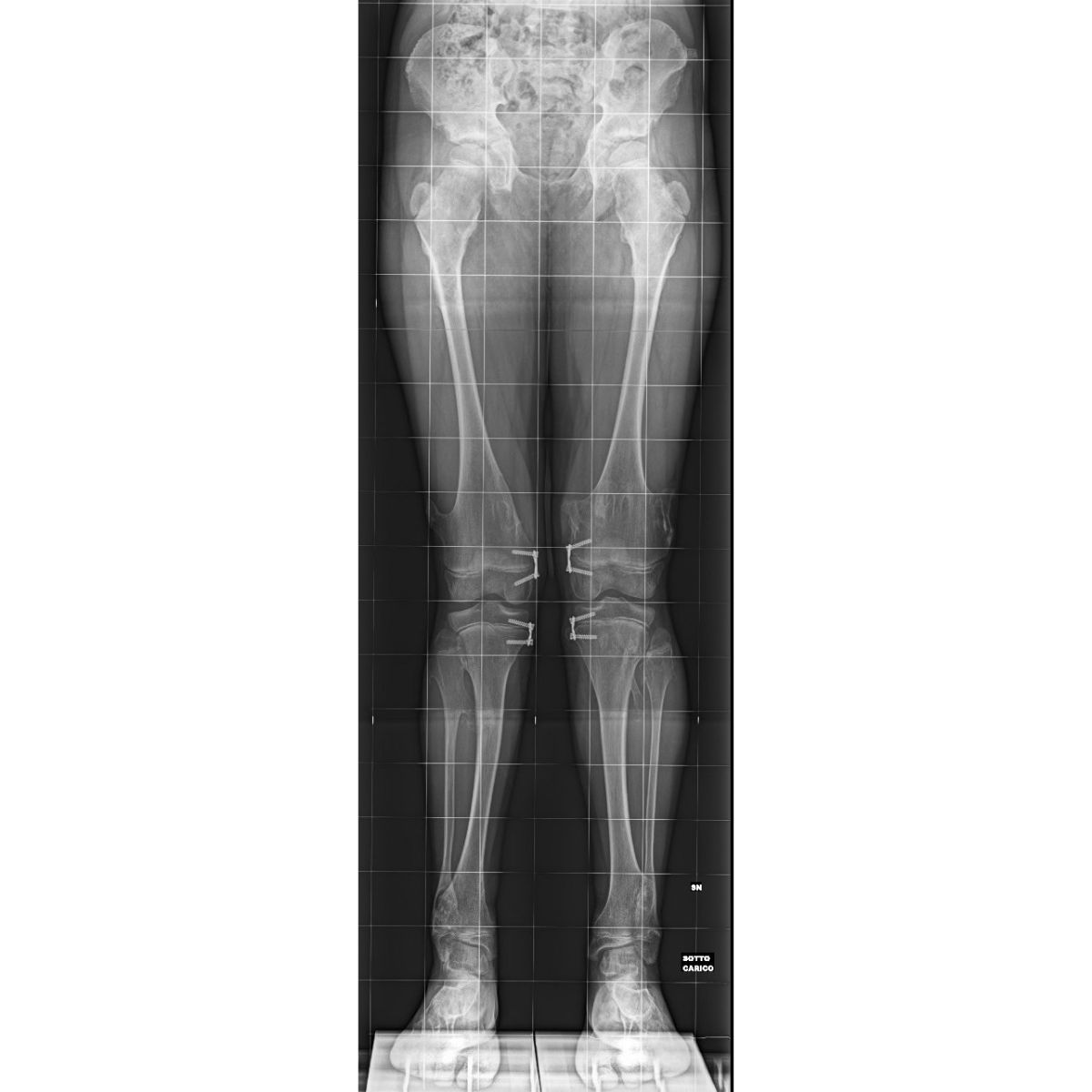
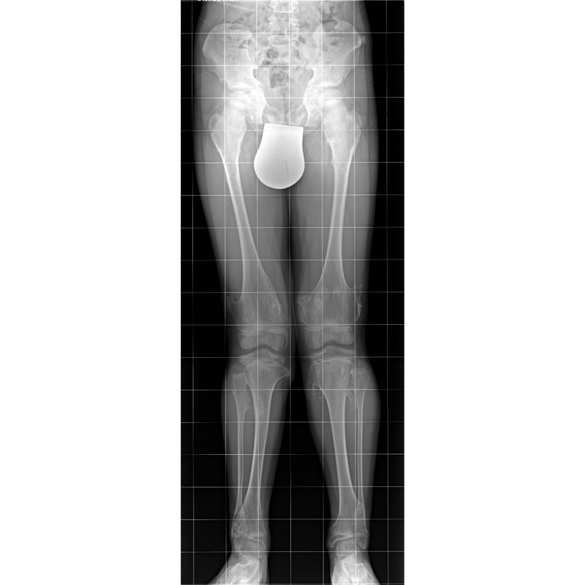
Full radiographs of the lower extremities (pangonogram) in the standing position
Measurement of the mechanical angles of the limb axes
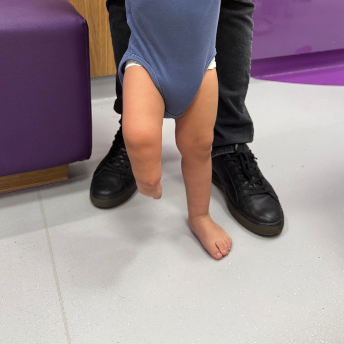
Assessment of Limb Length Discrepancy
Diagnosis of the cause (metabolic, post-traumatic, congenital, degenerative)
A personalized treatment plan tailored to the patient's age and the severity of the deformity
The global standard in the treatment of lower limb deformities
The Paley European Institute is one of the few centers in Europe offering a full range of treatments for lower limb deformities—using methods developed and refined by Dr. Dror Paley, a world leader in limb reconstructive surgery. Patients are treated by the most experienced specialists.
Treatment of lower limb deformities begins with a detailed analysis of the biomechanics of the entire musculoskeletal system, rather than just the joint where symptoms appear. This approach makes it possible to precisely identify the site of the deformity and plan the most effective treatment, rather than limiting treatment to symptom management.
In our treatment, we use, among other things:
The Method Guided Growth —a minimally invasive method for correcting limb alignment during a child’s growth period
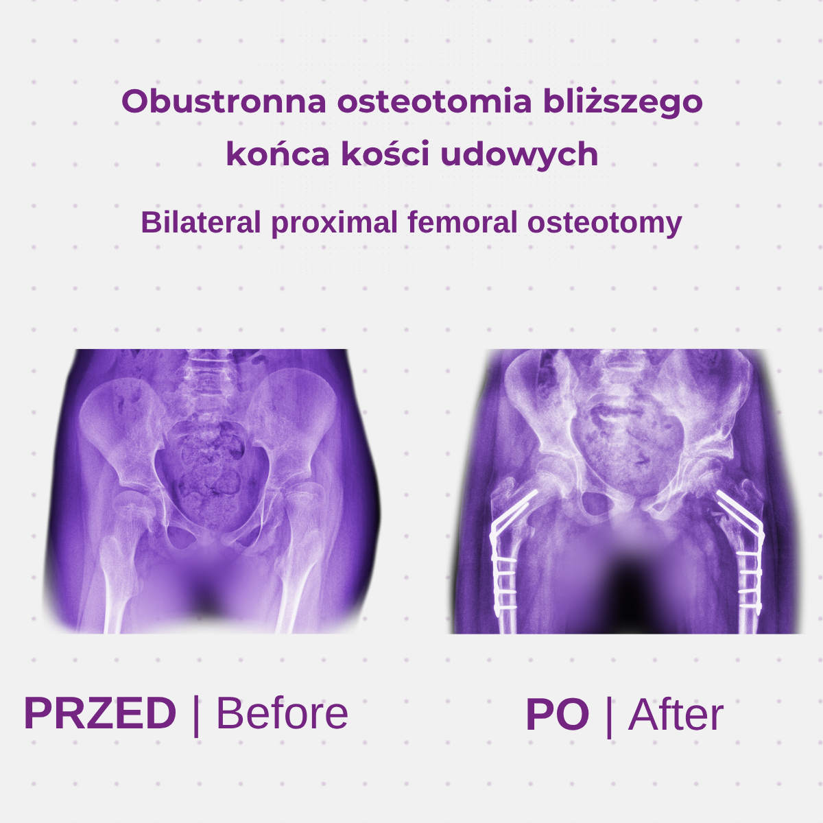
Precise corrective osteotomy of the femur or tibia
Modern techniques for the gradual lengthening of limbs
In advanced osteoarthritis— knee replacement
Physiological Development of the Limb Axis—When Is It Still Considered Normal?
Physiological knock-knees is a natural condition in newborns and infants, resulting from the fetal position. Over time, usually 6–12 months after the child begins walking, it transitions to physiological valgus, which stabilizes in children by age 7, reaching the angle typical for adults (approximately 6–7 degrees). If limb alignment abnormalities exceed these physiological limits, consultation with an orthopedist or physical therapist is necessary.
Key developmental milestones:
Newborns and infants – physiological knee valgus
6–12 months of walking – transition to physiological valgus
By age 7 – stabilization of the limb axis to values typical for adults
Outside the age-appropriate range – indication for specialized diagnosis
In treating children, the Paley European Institute uses the Guided Growth —a minimally invasive technique that temporarily blocks growth plates, allowing for gradual, natural correction of limb alignment during growth, without the need for extensive procedures in the future.
When should you see a specialist?
Infants and young children (up to about 2 years of age)
Mild bowlegs are a natural stage of development. Concerns should arise if the deformity worsens instead of gradually improving, if there is asymmetry between the legs, or if it is accompanied by short stature or other developmental abnormalities.
Children aged 2–7 years
This is a period of physiological valgus, which should gradually decrease. Persistent or worsening valgus, especially when unilateral, requires orthopedic evaluation, as it may indicate Blount’s disease or another pathological condition.
Children over 7 years of age and adolescents
After this age, the limb axis should be stabilized. Persistent deformity, knee pain, uneven limb length, or noticeable asymmetry in gait are indications for an urgent specialist consultation.
Adults
Limb alignment deformities in adults are often associated with pain, limited mobility, and progressive degenerative joint changes. It is advisable to consult a specialist if the deformity worsens, is accompanied by pain, or if previous treatment has not produced the expected results.
How do we treat lower limb deformities?
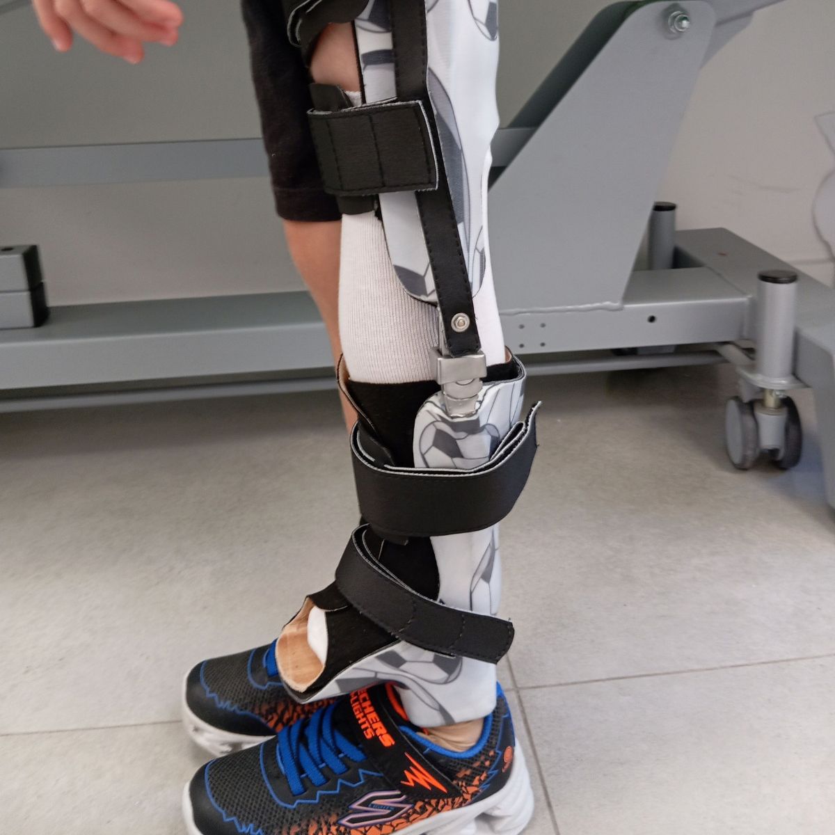
A comprehensive approach to treating limb alignment deformities is based on precise diagnosis, identification of the cause, and tailoring the treatment method to the patient’s age and the severity of the condition. The team at the Paley European Institute combines modern surgical techniques with individually tailored rehabilitation so that patients can regain normal limb function.
Diagnostics and Planning
Objective: To precisely determine the cause and extent of the deformity
Full radiographs of the limbs, measurement of mechanical axis angles
Eligibility for Conservative or Surgical Treatment
Developing a personalized treatment strategy tailored to the patient's age
Guided Growth (Minimally Invasive Treatment for Children)
Goal: Controlled correction of the axis during the growth period
Temporary closure of the growth plate using a figure-eight plate
Implantation through two small incisions – minimal risk of scarring
It helps avoid more invasive procedures in the future
Corrective Osteotomy (Adolescents and Adults)
Objective: Surgical correction of a fixed axial deformity
Used when growth potential has been exhausted or the deformity is advanced
It can be combined with gradual limb lengthening
In advanced osteoarthritis—total knee replacement is being considered
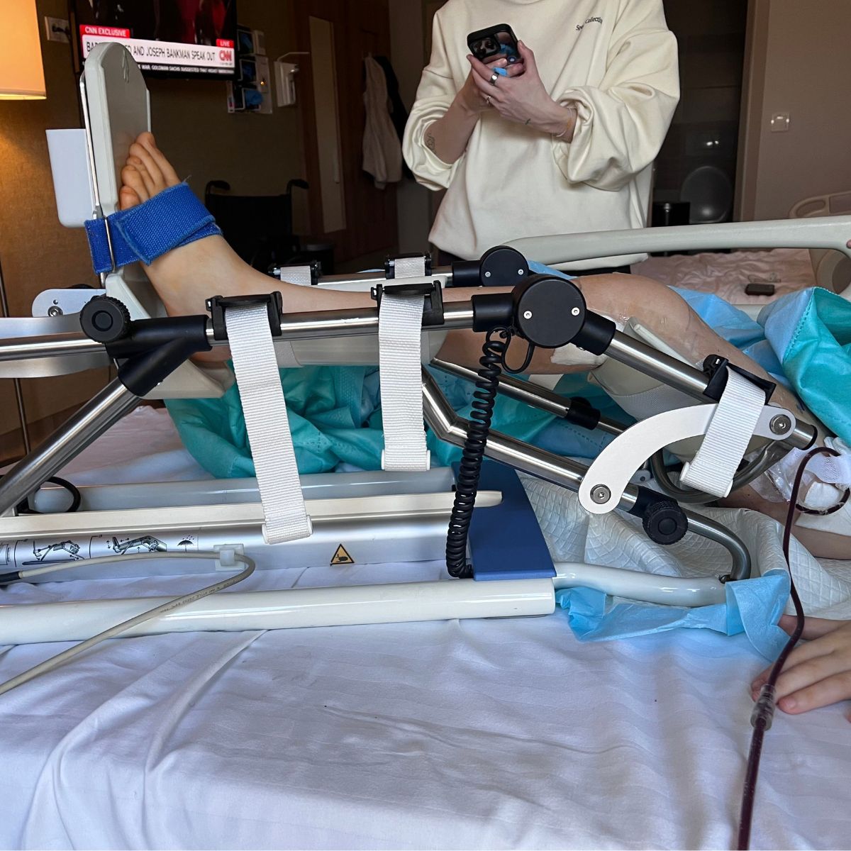
Rehabilitation and Long-Term Care
Goal: Full recovery of physical fitness and proper gait pattern
A personalized physical therapy program
Regular radiographic monitoring of the progress of the correction
Long-term monitoring until the end of growth (in children) or full recovery (in adults)
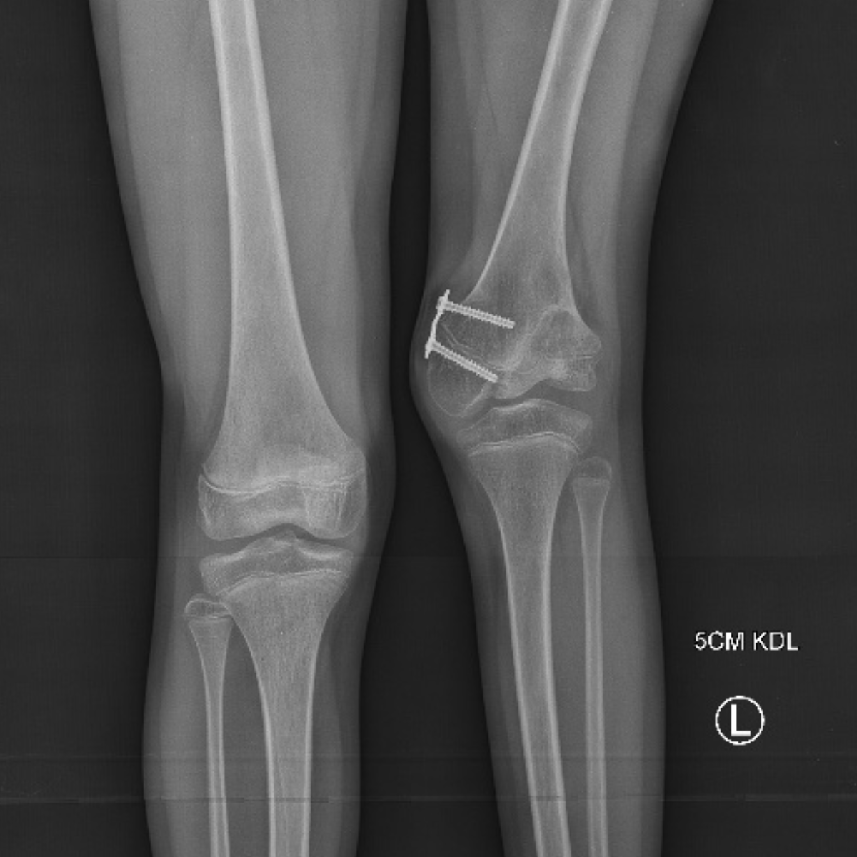
Bowlegs (Genu Valgum)
Bowlegs is a deformity of the lower limb in which the knees are positioned unnaturally close together, while the feet remain spread apart—when viewed from the front, the legs form an “X” shape. Mild bowlegs is a physiological stage of development in children between the ages of 2 and 7 and usually resolves on its own. Persistent or worsening bowlegs after age 7 require specialized evaluation.
Symptoms:
– Visible inward turning of the knees when the feet are spread apart
– Uneven wear on shoes
– Pain in the front or medial part of the knee
– Instability while running and during physical activity
– Fatigue in the lower limbs after exercise
Treatment:
In growing children, the treatment of choice is the Guided Growth method—a minimally invasive procedure involving the implantation of an octagonal plate that temporarily blocks one side of the growth plate, allowing for gradual, natural correction of the limb’s axis as the child continues to grow.
Management in adolescents and adults:
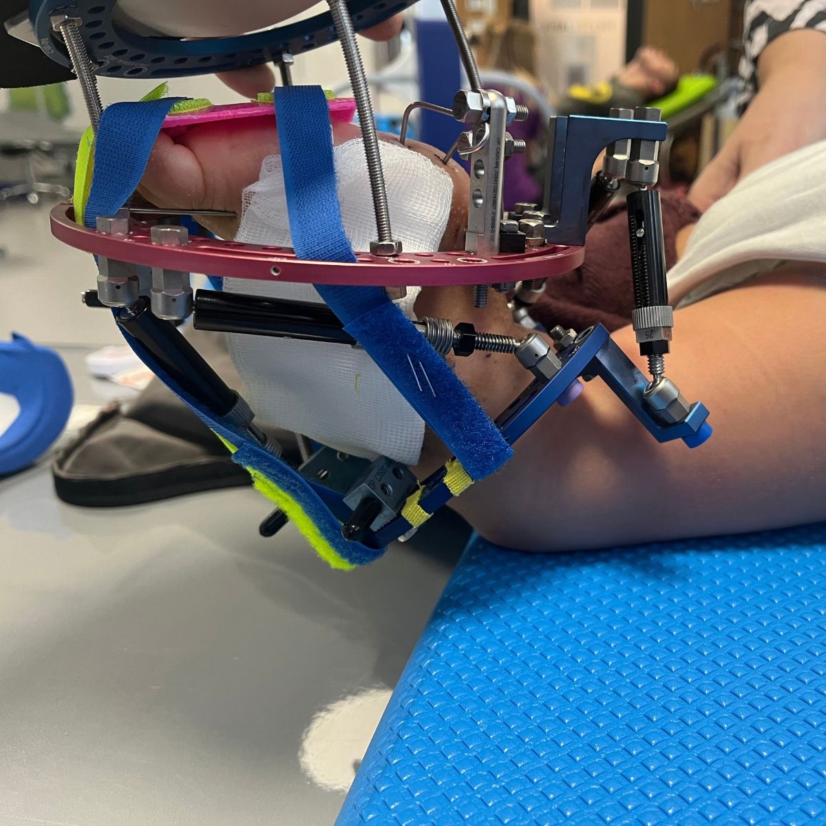
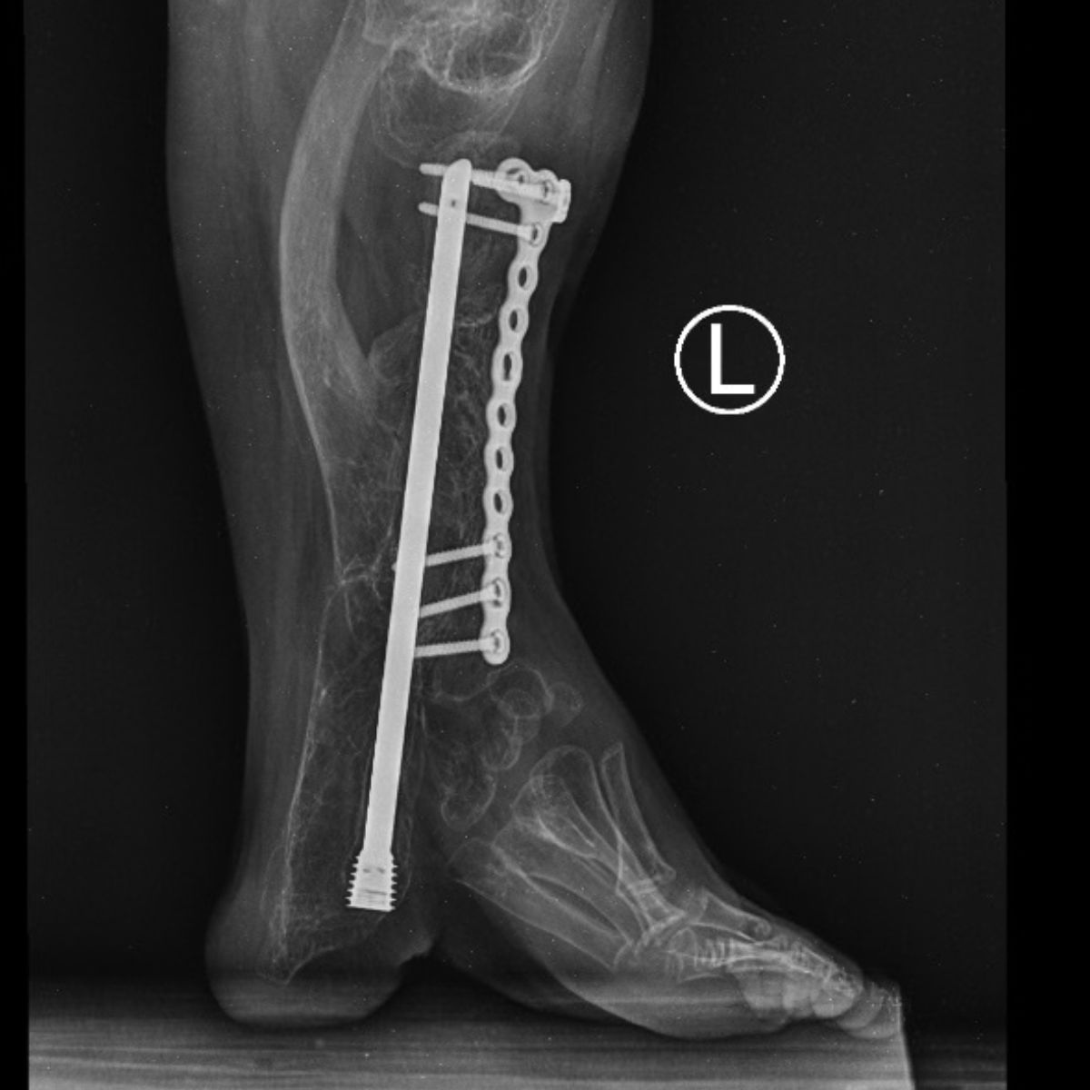
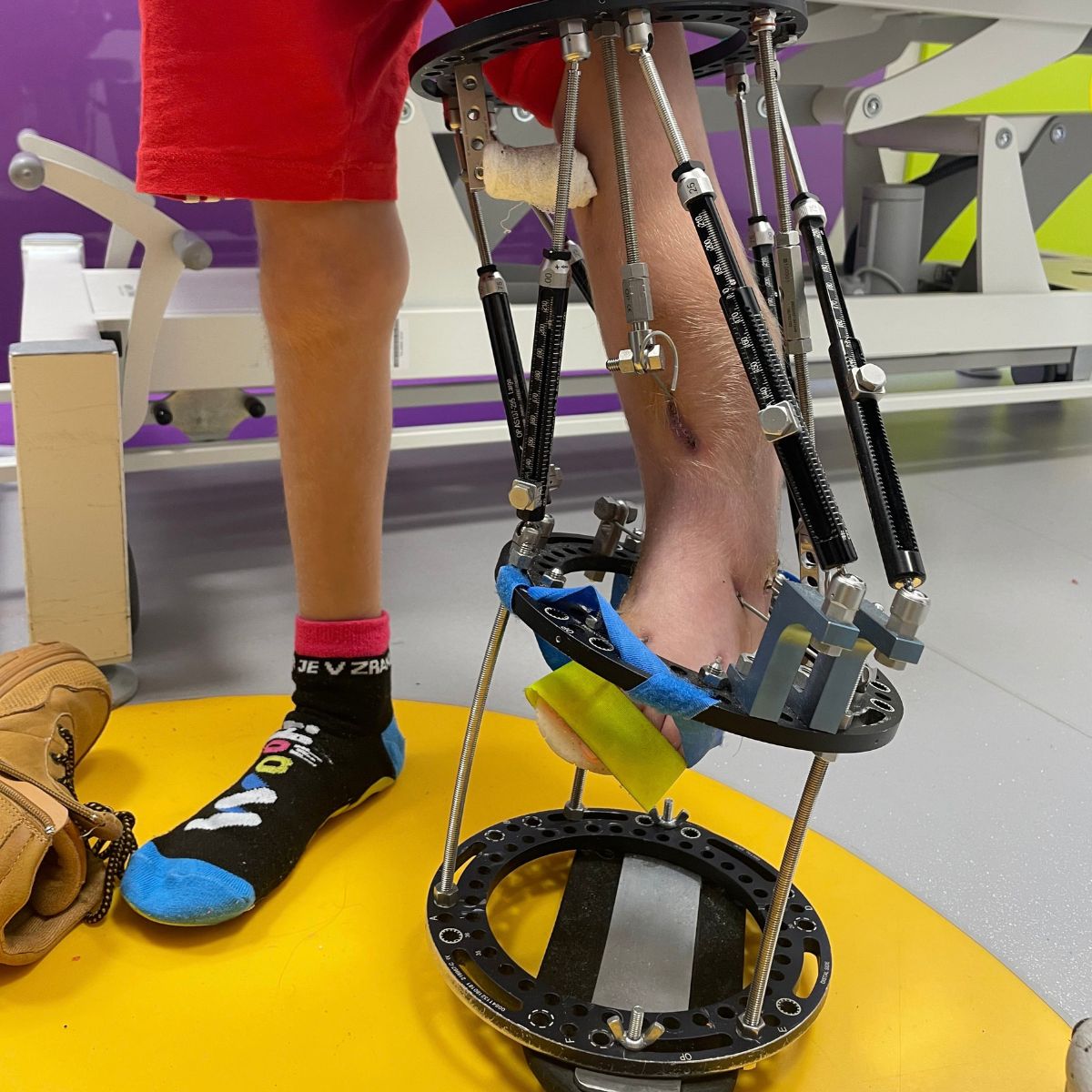
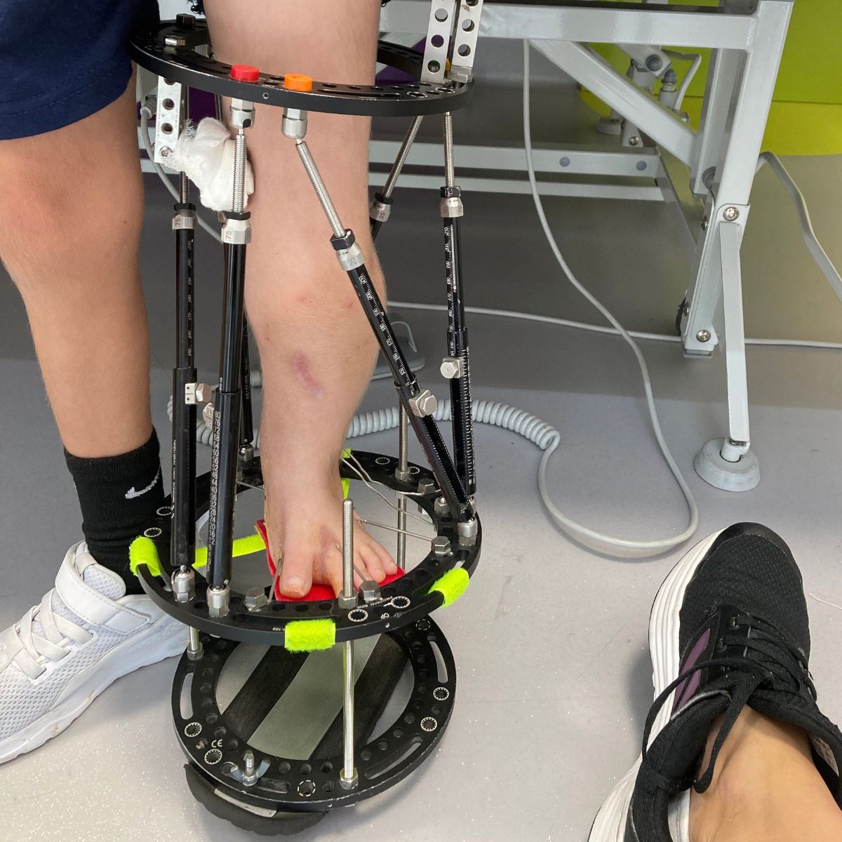
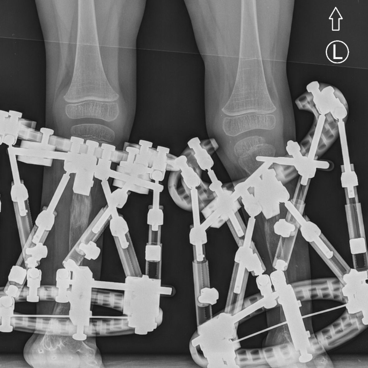
When growth potential has been exhausted and the deformity has become permanent, a corrective osteotomy is performed—a planned cut and realignment of the bone to its proper axis, stabilized using plates or external fixators.
Rehabilitation:
The physical therapy program is tailored to each individual and includes restoring range of motion, strengthening the muscles that stabilize the knee, and a gradual return to full physical activity under the supervision of the therapeutic team.
Proper treatment of knock-knees helps prevent secondary degenerative changes in the knee joint and ensures that patients can comfortably regain their mobility.
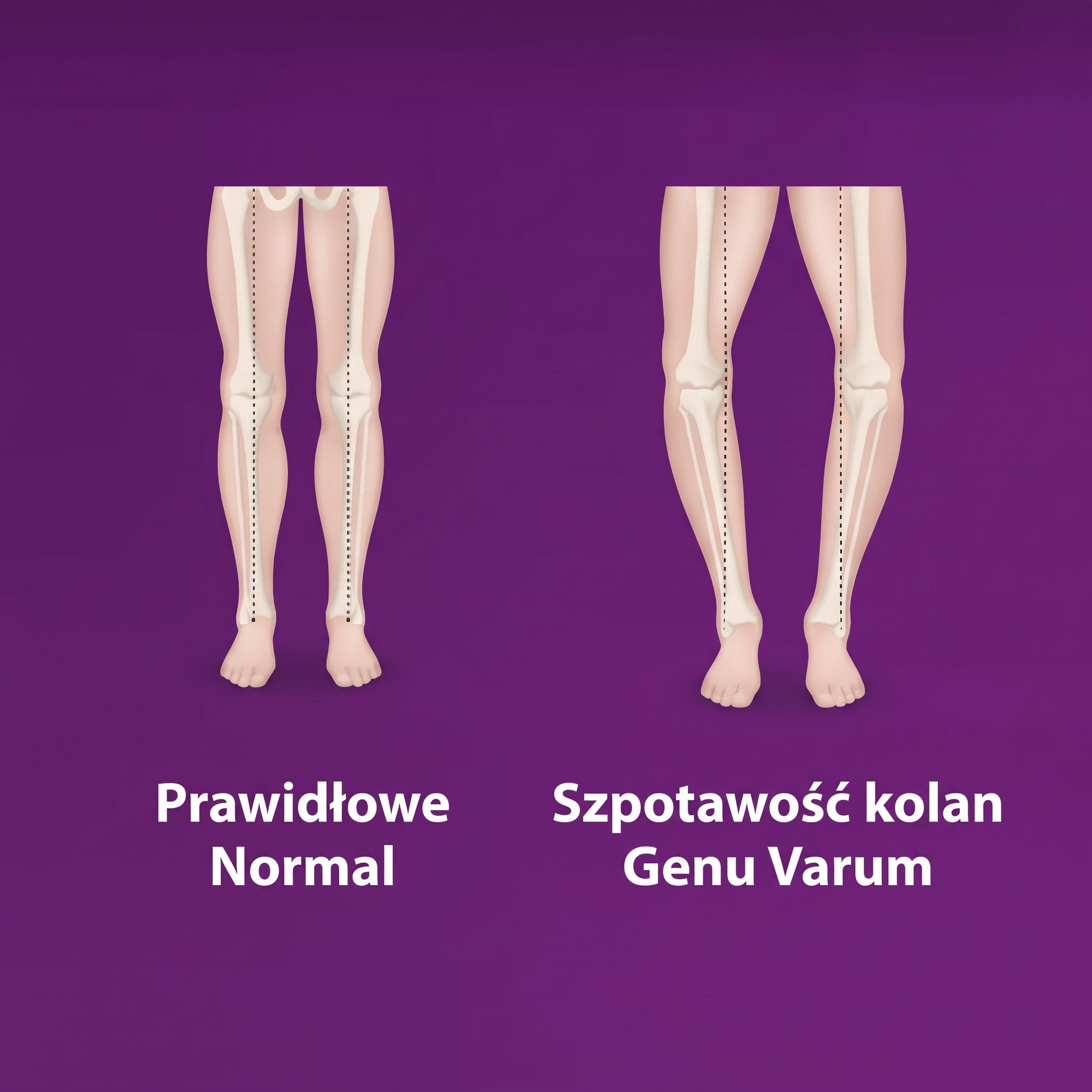
Bowlegs (Genu Varum)
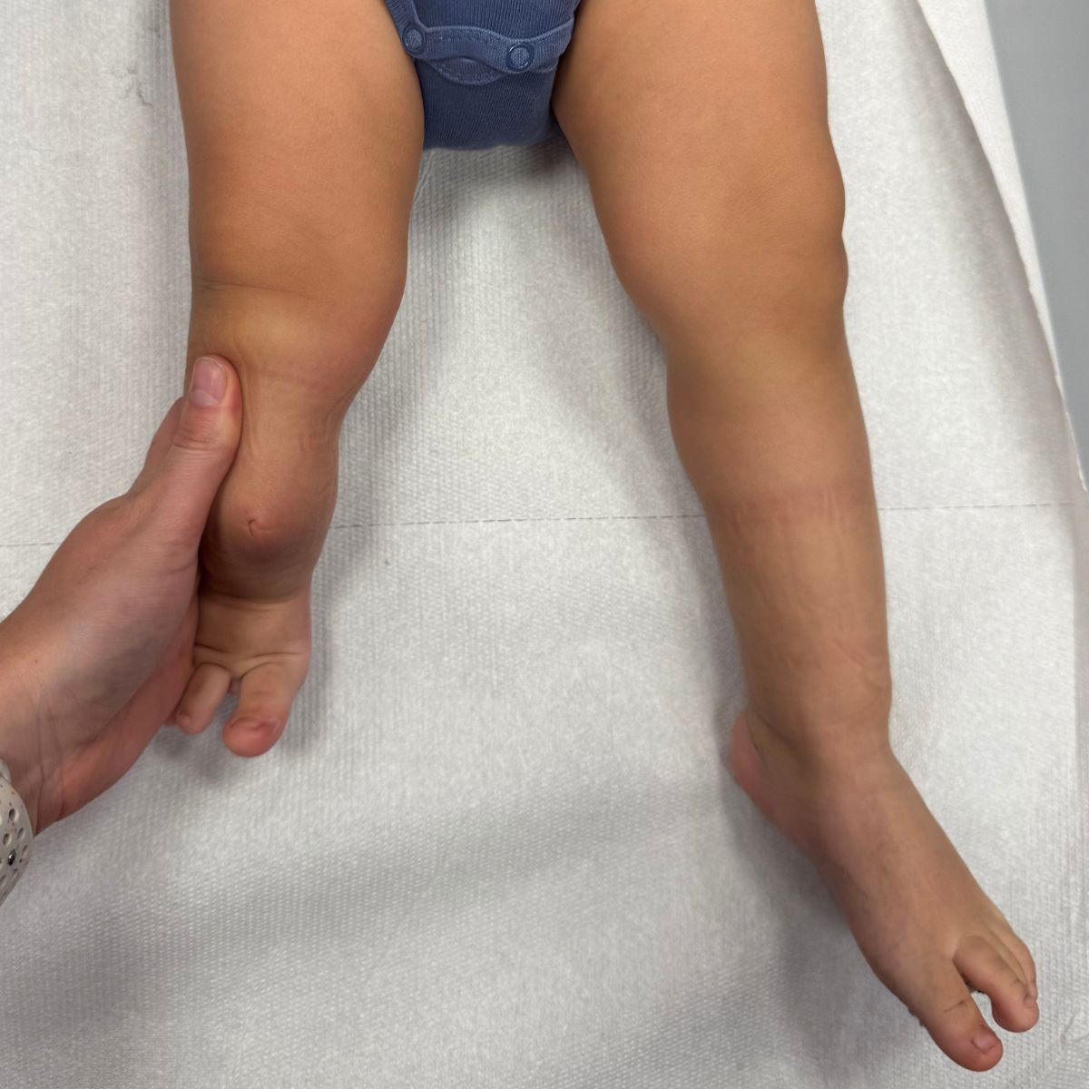
Bowlegs is a deformity of the lower limb in which the knees are unnaturally far apart, while the feet remain close together—viewed from the front, the legs form an “O” shape. Mild bowlegs are normal in newborns and infants due to fetal positioning and usually resolve on their own by age 2, as the child begins to walk. Persistent or worsening bowlegs after this age, especially if unilateral or asymmetrical, requires urgent evaluation—it may indicate Blount’s disease, rickets, or another underlying pathological condition.
Symptoms:
– Visible splaying of the knees while the feet are together
– A waddling, “duck-like” gait
– Pain on the medial or lateral side of the knee
– Knee instability when bearing weight
– Uneven wear on the outer edge of shoes
– Worsening of the deformity instead of its gradual resolution
Treatment:
In young children with persistent pathological bowlegs (e.g., due to Blount’s disease), the Guided Growth method is used—a minimally invasive, temporary blocking of the outer side of the growth plate with a figure-eight plate, which allows for gradual straightening of the limb’s axis as the child grows naturally.
Management in adolescents and adults:
Once growth has ceased or in cases of advanced, established deformity, a corrective osteotomy of the tibia or femur may be necessary to restore the limb’s proper mechanical axis and relieve pressure on the medial part of the knee joint, which is most susceptible to degenerative changes.
Rehabilitation:
Physical therapy focuses on strengthening the muscles that stabilize the knee and hip, correcting gait patterns, and gradually increasing the load, with regular radiographic monitoring of progress.
Early diagnosis and treatment of bowlegs, particularly in cases of Blount's disease, are crucial for preventing permanent damage to the articular cartilage and premature degenerative changes.
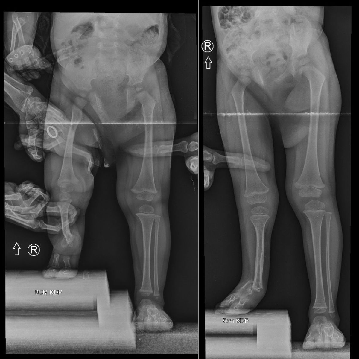
Limb Length Discrepancy (LLD)
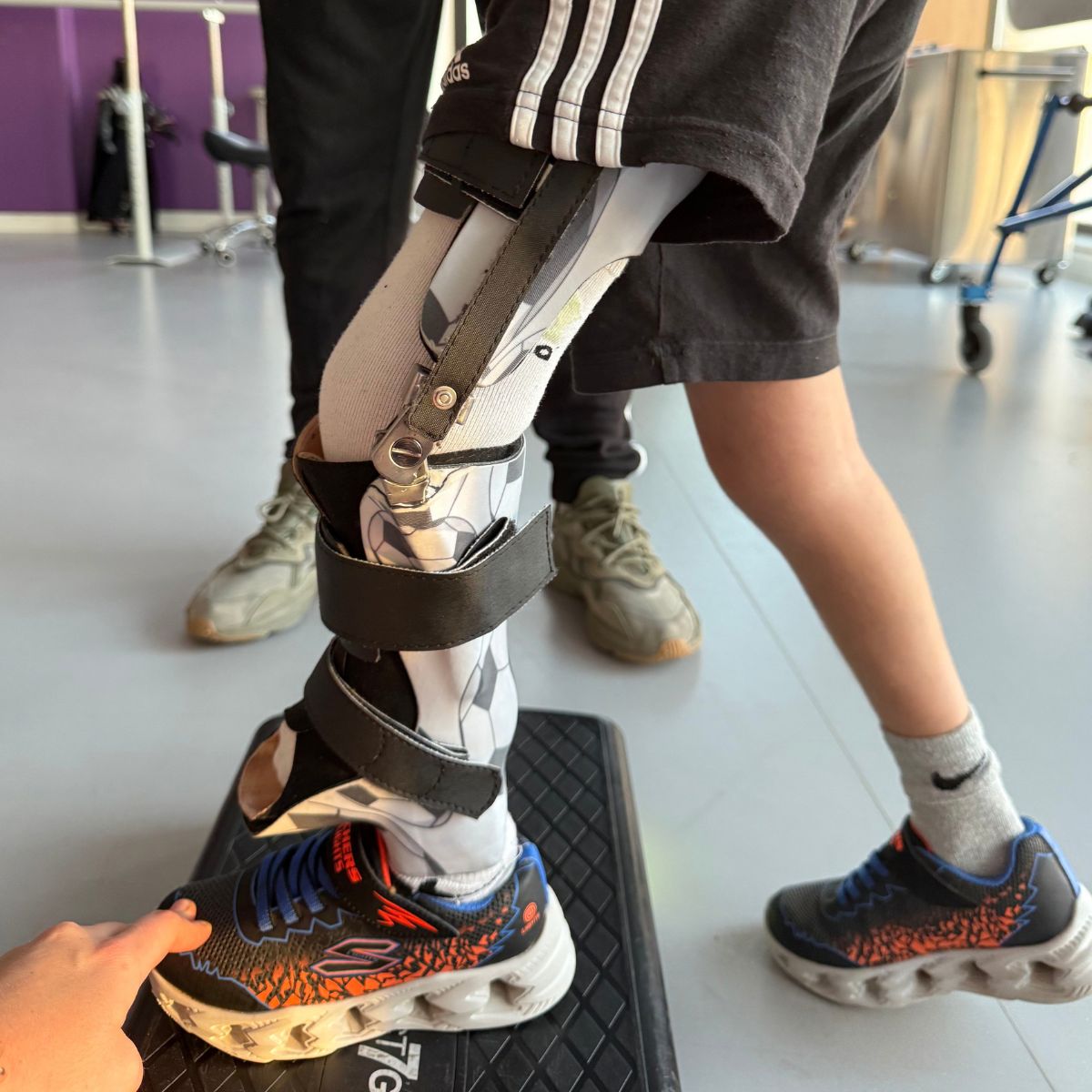
Lower extremity length discrepancy is a difference in leg length that may be congenital or acquired. In children and adolescents, it most often results from paralytic disorders (e.g., cerebral palsy, polio), injuries to the bone growth plate (trauma, inflammation, neoplastic tumor), congenital syndromes (osteochondrodysplasia, proximal femoral hypoplasia, fibular hemimelia, Silver-Russell syndrome), or hyperplastic and vascular syndromes. In adults, this discrepancy is often the result of previous growth disorders, injuries, or fractures.
Symptoms
– Visible leg-length discrepancy
– Compensatory alignment of the pelvis and spine
– Limping or an abnormal gait pattern
– Pain in the hips, knees, or lumbar spine
– Increased risk of degenerative changes in the hip joint on the side of the longer limb
Treatment:
The choice of treatment depends on the patient’s age, the cause, and the expected difference in leg length in adulthood. In children who are still growing, Guided Growth methods are used (temporary or permanent locking of the growth plate in the longer limb—epiphysiodesis), which allow leg length to be equalized as the child grows naturally.
Management of Significant Length Discrepancies:
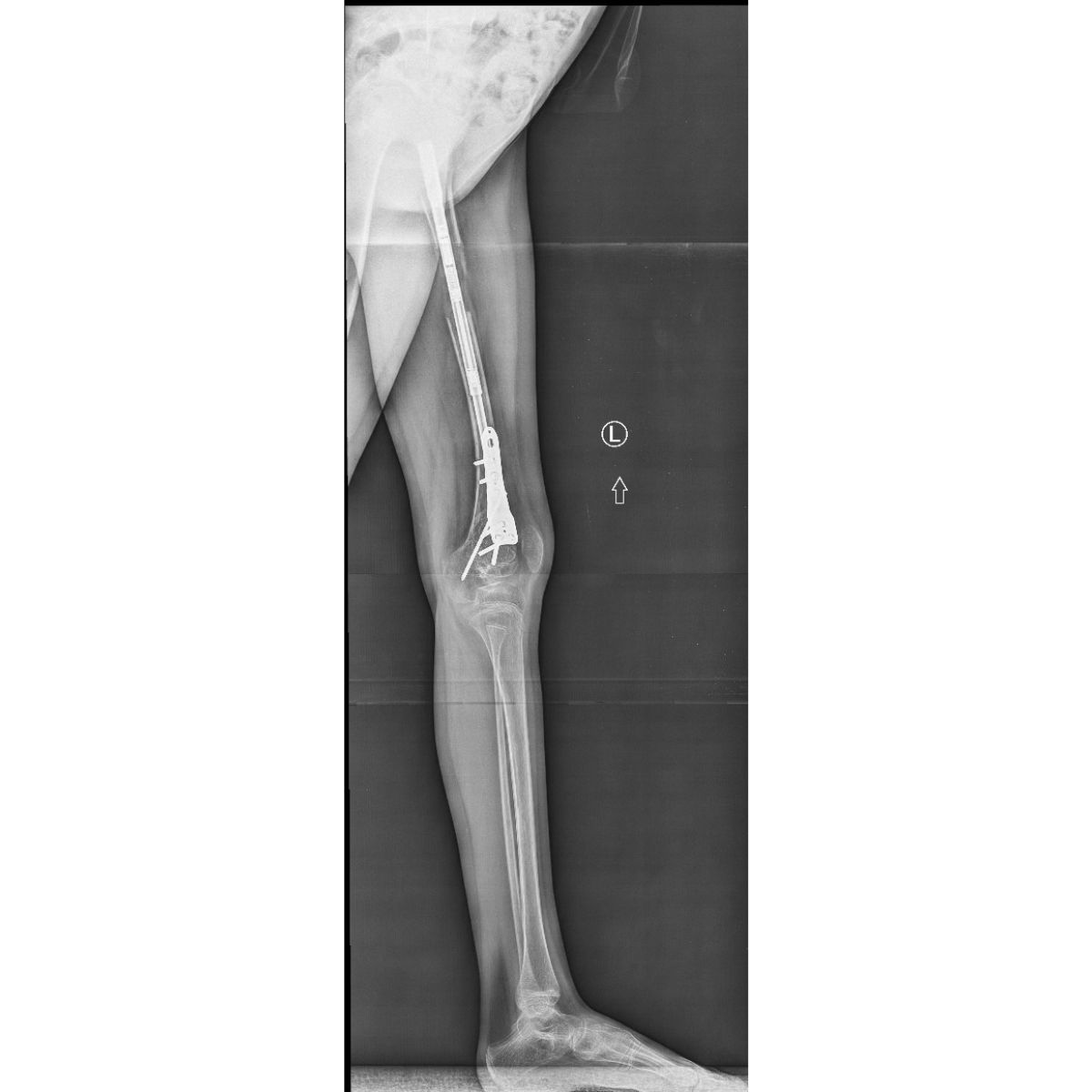
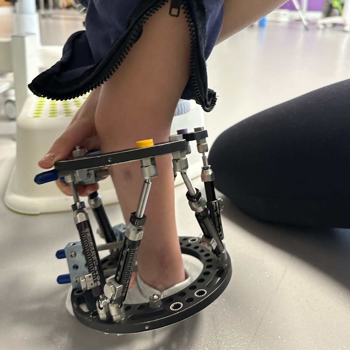
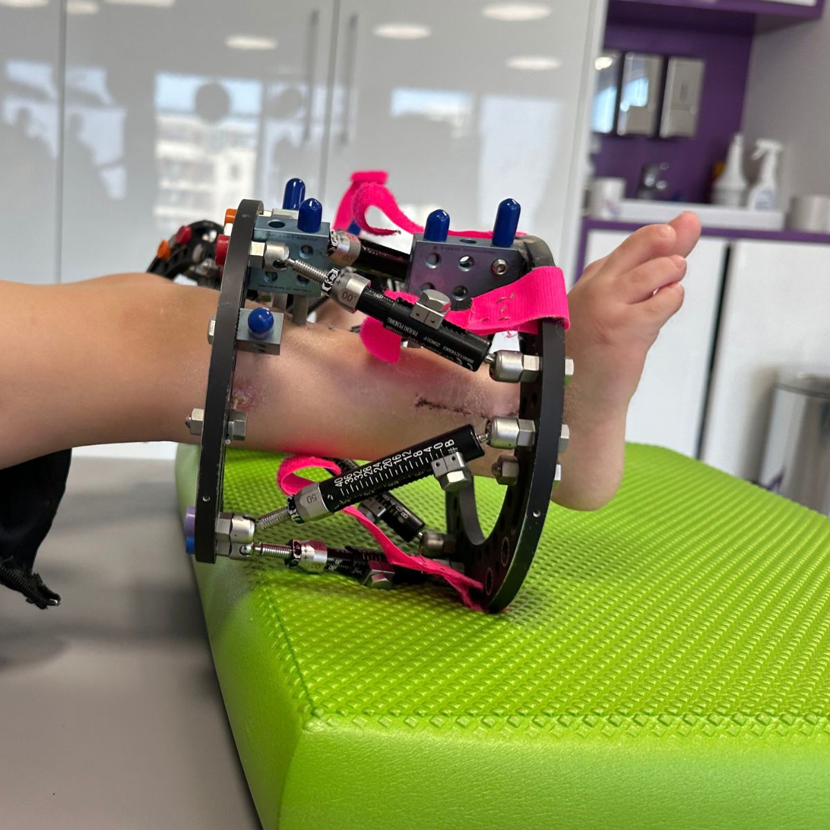
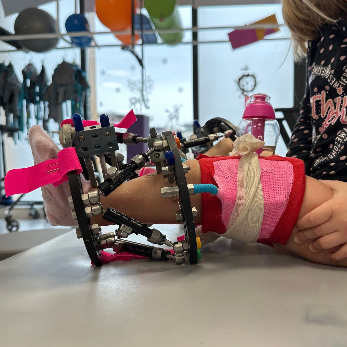
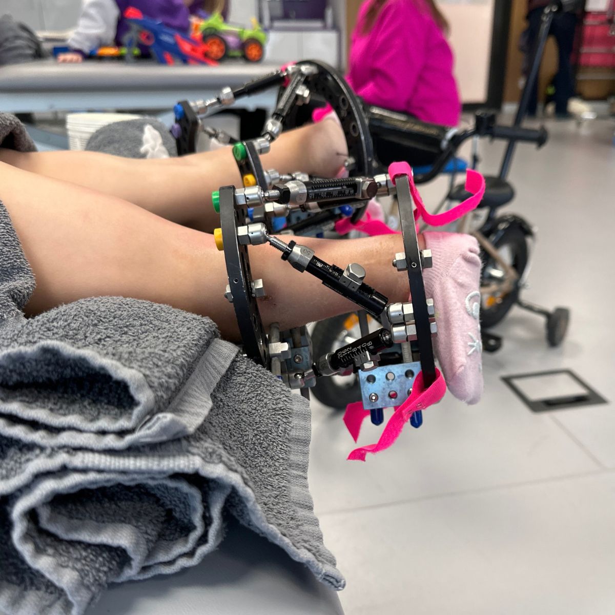
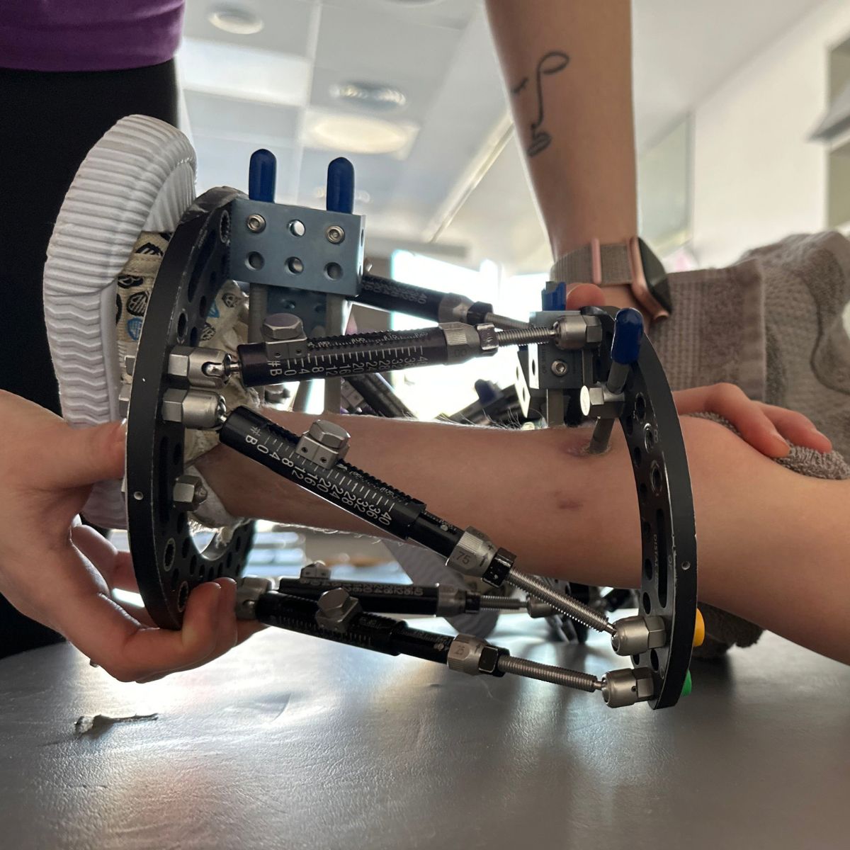
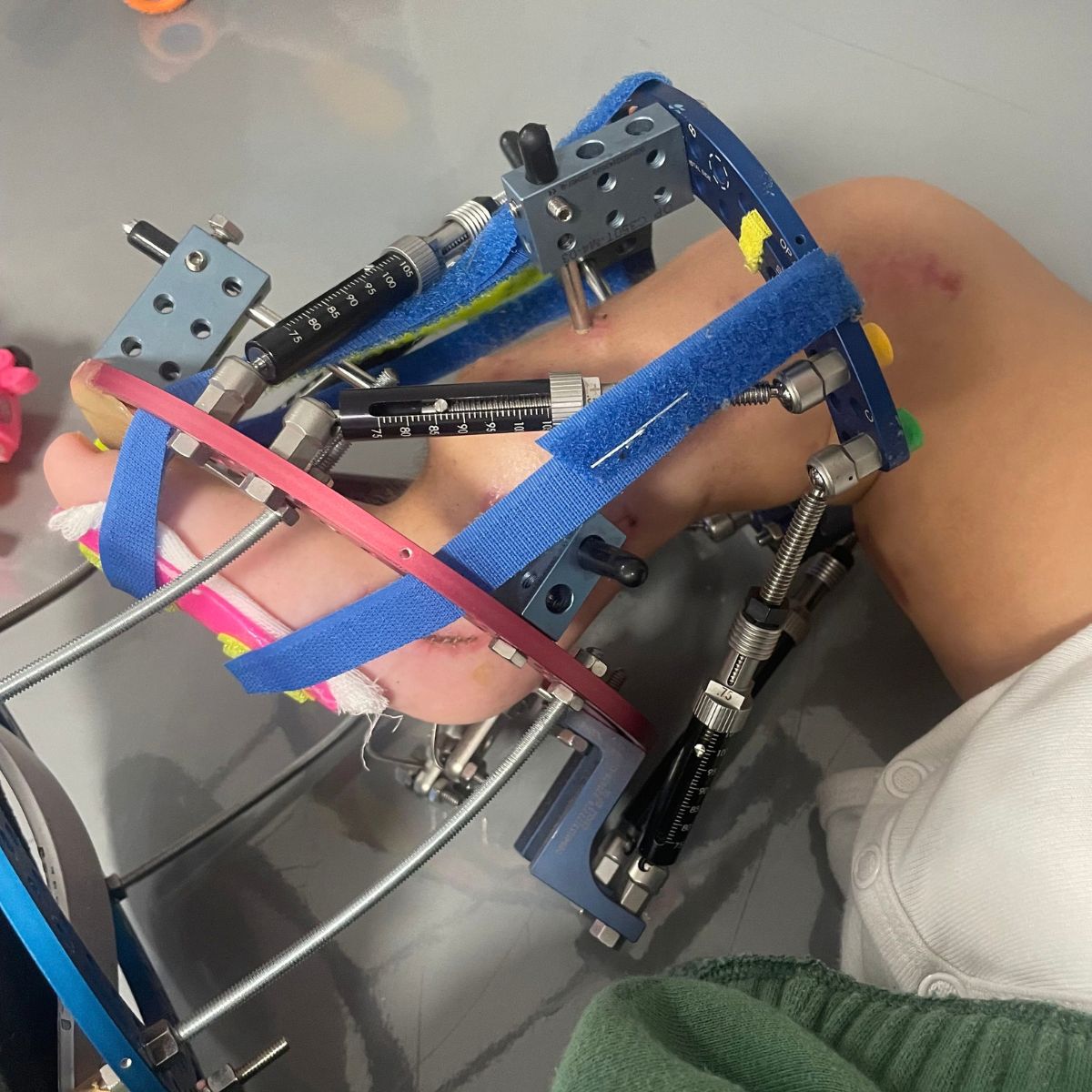
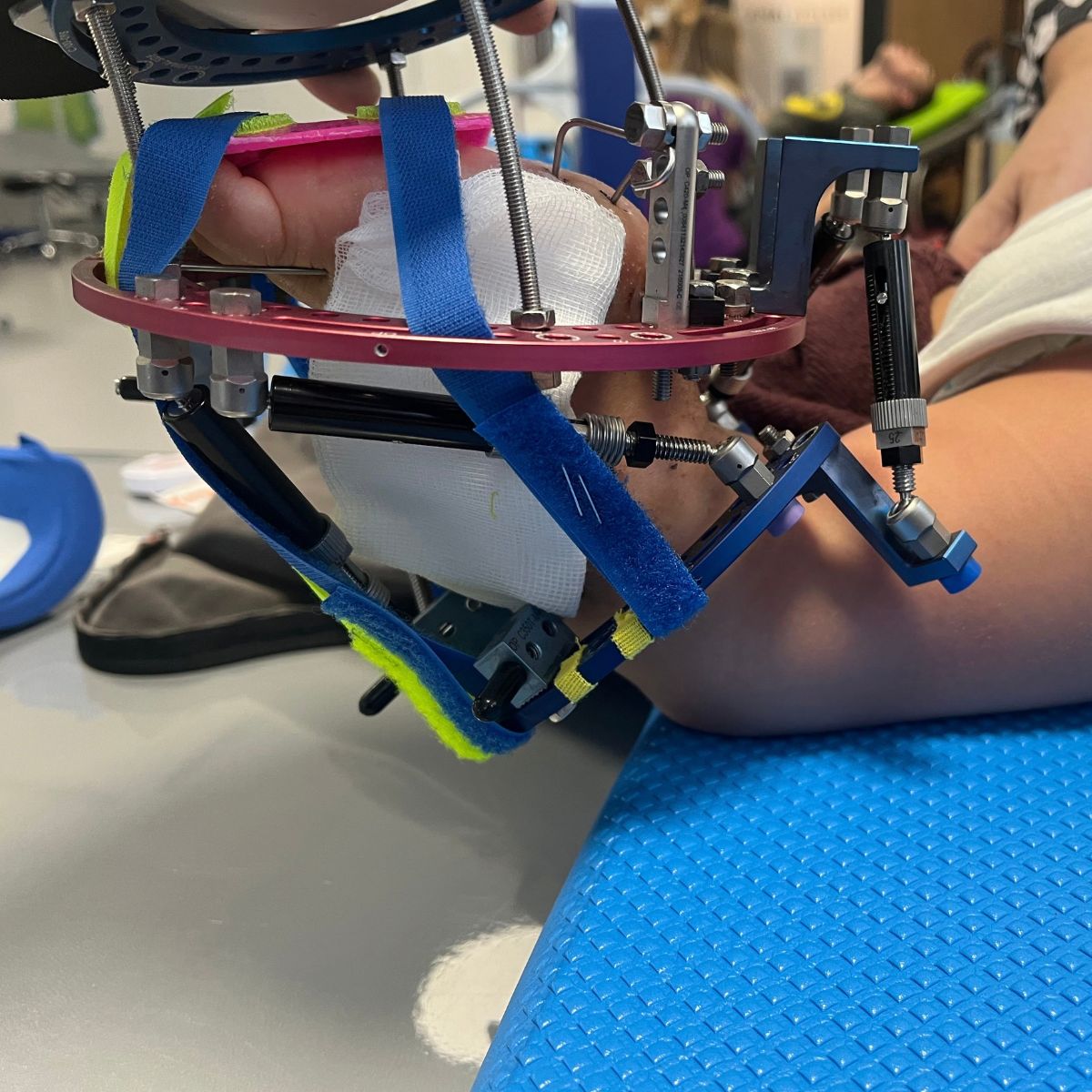
In cases of significant length discrepancies, in both children and adults, gradual limb lengthening is performed using modern intramedullary nails or external fixators, which allow for precise, controlled correction.
Rehabilitation:
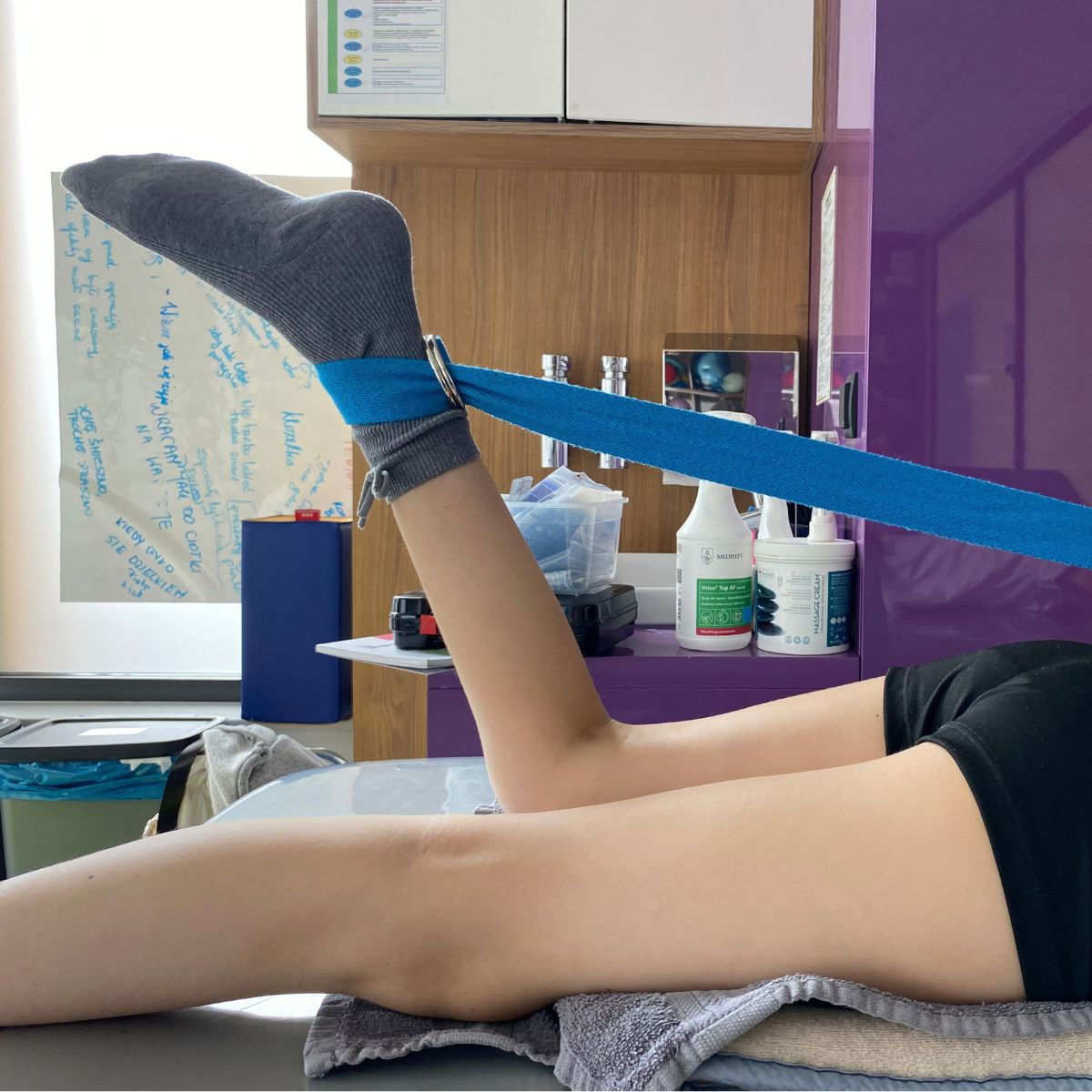
The process of lengthening and equalizing limb lengths requires long-term, regular physical therapy to support bone regeneration and maintain a full range of motion in the adjacent joints, conducted under the constant supervision of a team of specialists.
Comprehensive diagnosis of the cause of limb length discrepancy and appropriately timed intervention allow for permanent correction and help prevent secondary postural disorders.
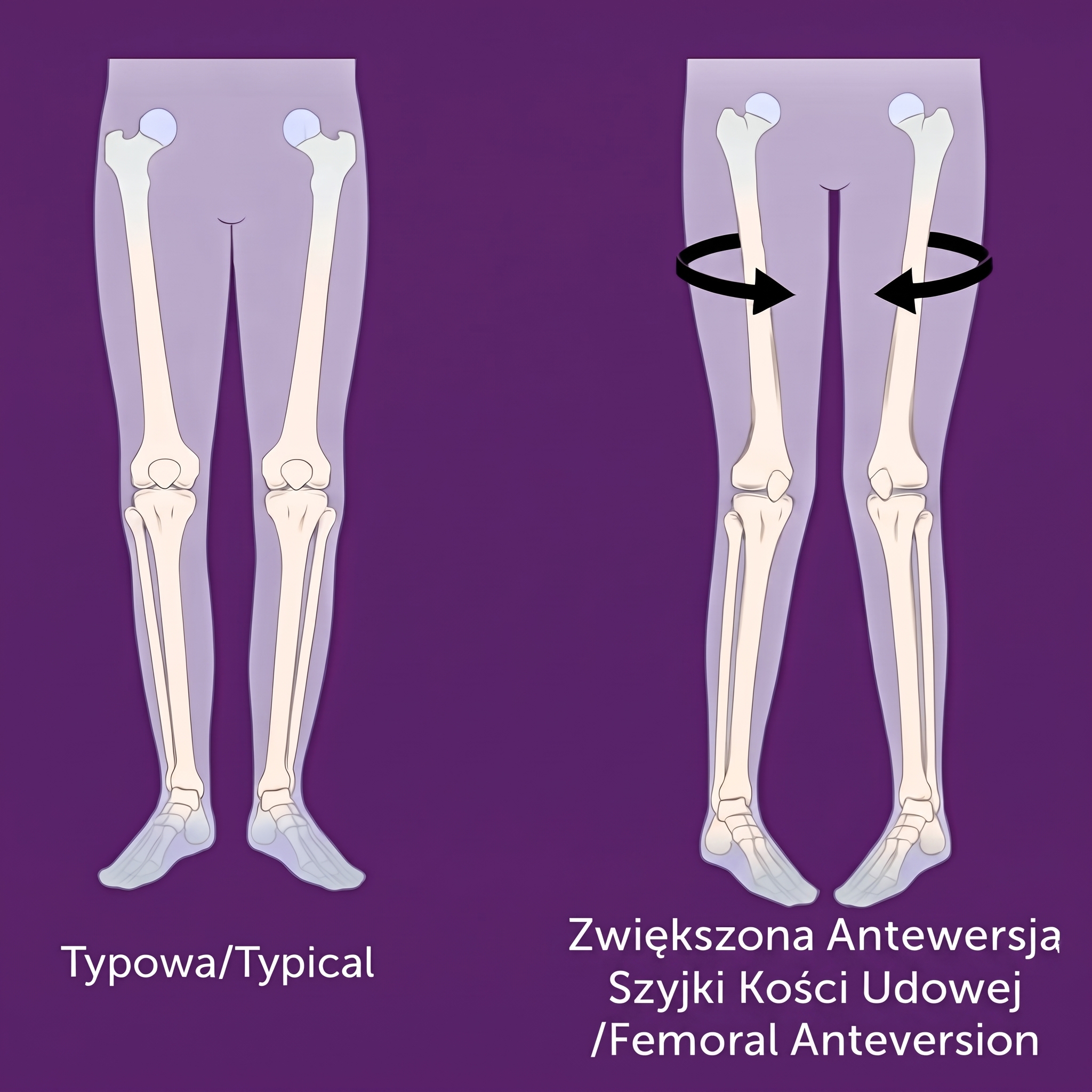
Increased Femoral Anteversion
Femoral neck anteversion is a natural forward rotation of the proximal part of the femur relative to its shaft. Increased anteversion refers to an excessive degree of this rotation, which causes a characteristic “inward” alignment of the knees and feet during walking. This condition is often observed in preschool-aged children and usually resolves on its own with age; however, in some cases, it persists and requires evaluation by a specialist.
Symptoms:
– “In-toeing” gait—feet and knees turned medially
– Frequent stumbling and an unsteady gait
– Preference for sitting in a “W” position (knees bent, feet turned outward)
– A feeling of tiredness in the legs after prolonged exertion
– In older children and adolescents—pain in the front of the knee associated with abnormal patellar tracking
Treatment:
In most children, increased anteversion resolves spontaneously by the age of 8–10 and does not require intervention—observation is recommended, along with supportive physical therapy if necessary. In cases that persist beyond this age and cause significant functional limitations or pain, surgical treatment is considered.
Surgical management:
In selected cases, a derotational osteotomy of the femur is performed—a planned cut in the bone to correct its rotation to the proper alignment, with internal fixation to ensure proper union.
Rehabilitation:
Postoperative physical therapy involves the gradual restoration of range of motion in the hip and knee joints, gait retraining, and strengthening the muscles that stabilize the pelvis and lower limbs.
A proper assessment of whether anteversion falls within physiological limits makes it possible to avoid unnecessary interventions in most children, while ensuring effective treatment for those in whom the deformity becomes permanent.
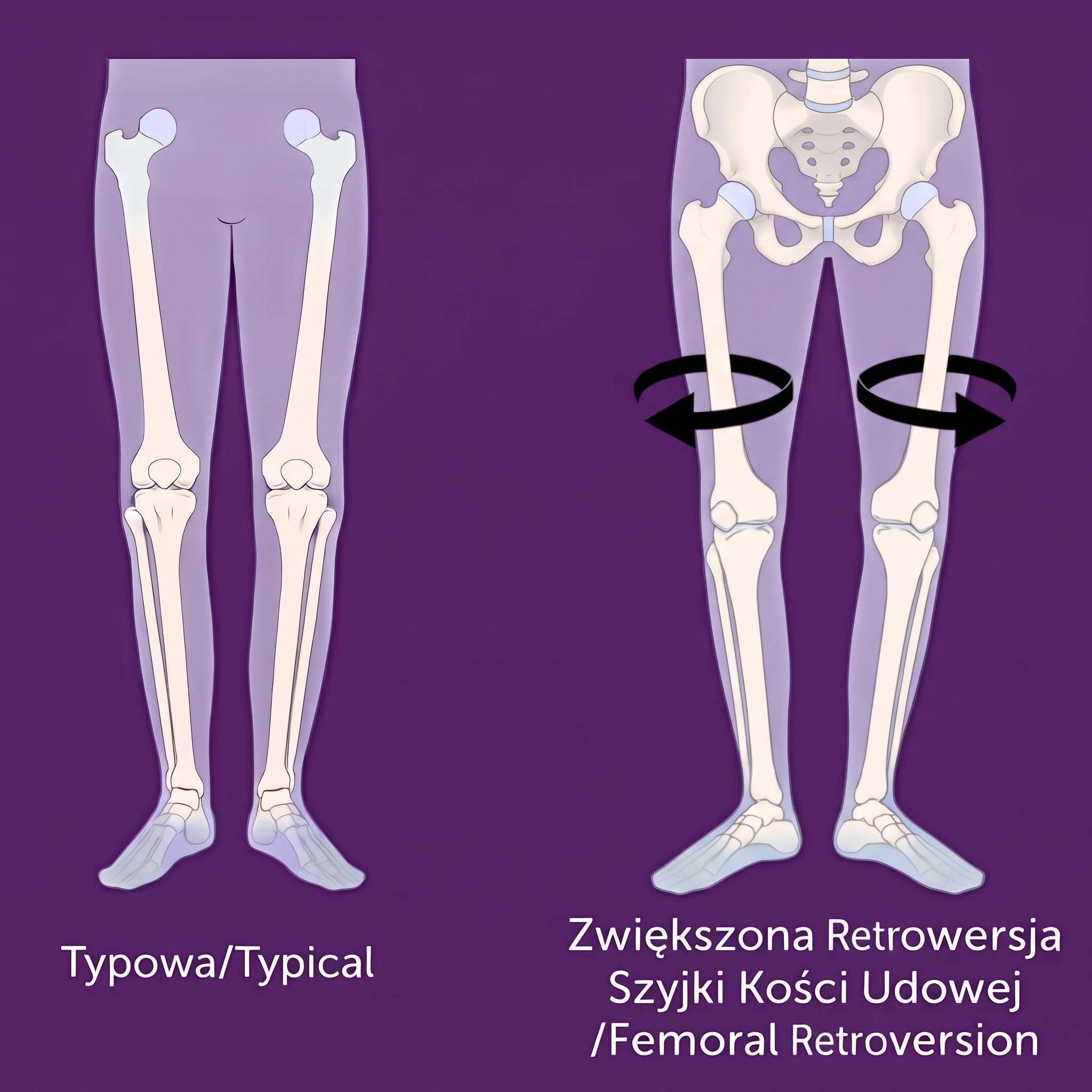
Femoral Retroversion, Nonunion, and Tibial Torsion
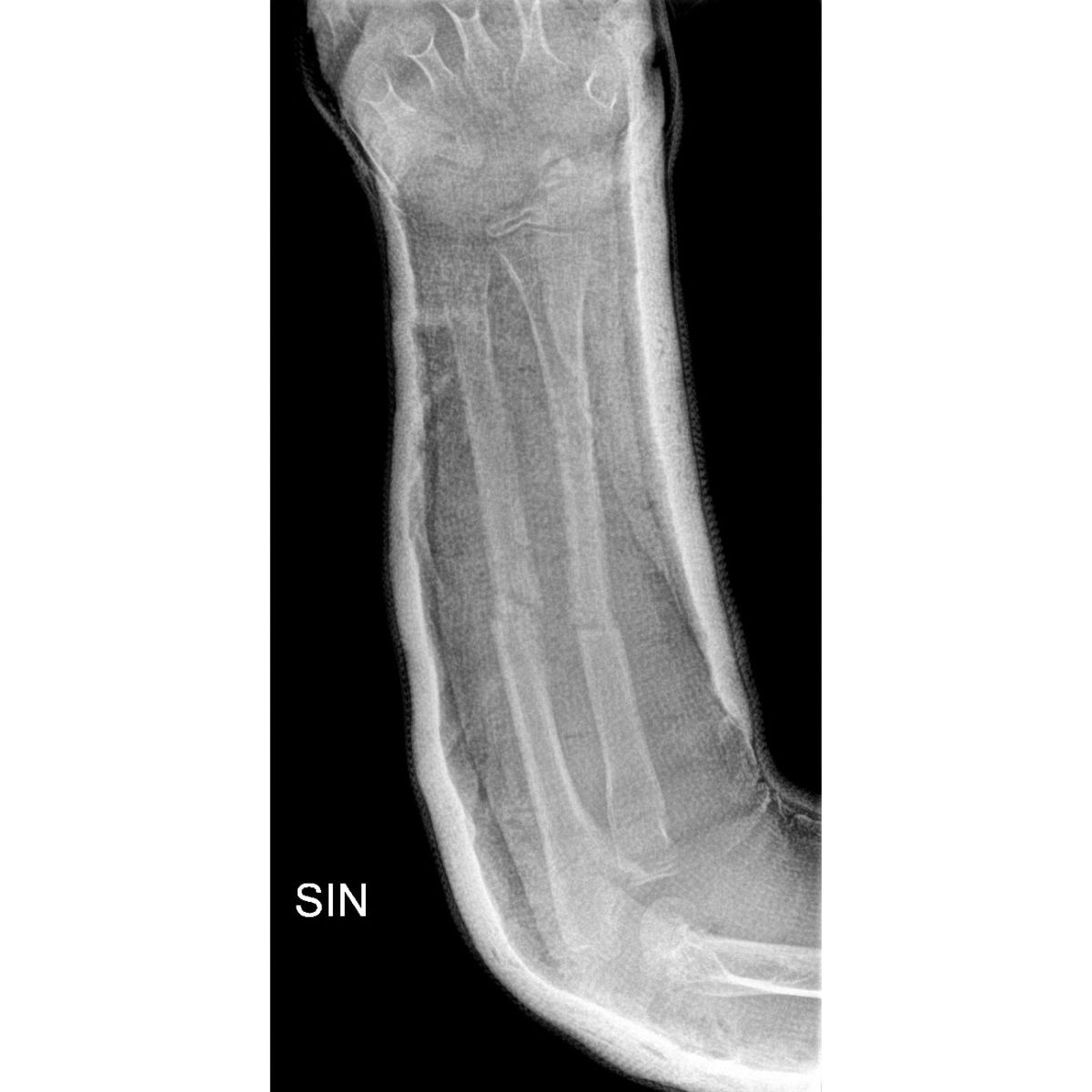
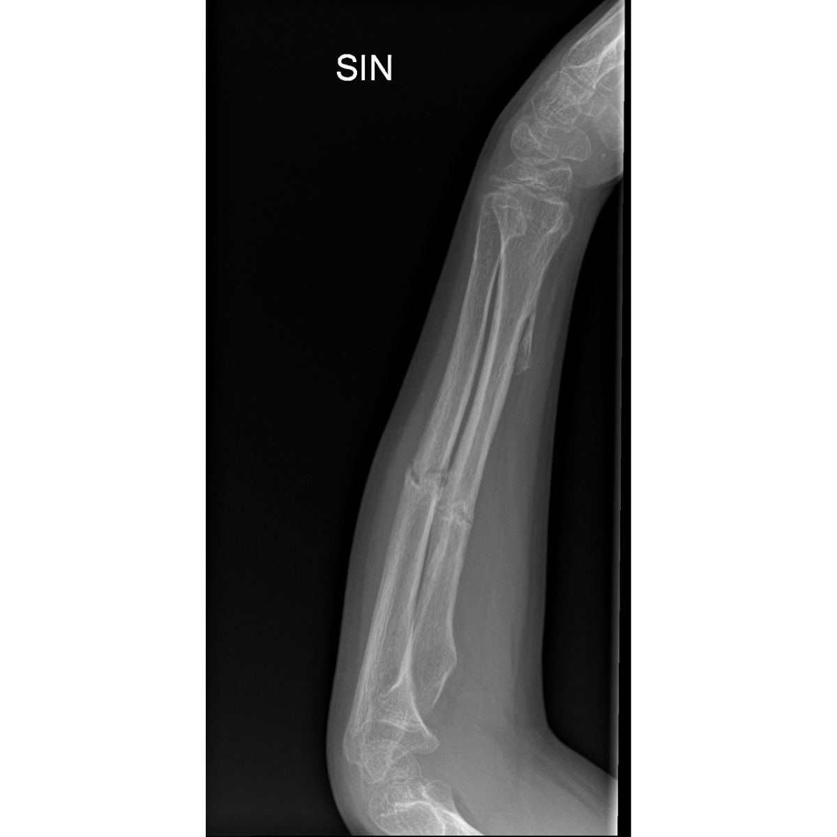
It is a group of complex rotational and structural disorders of the lower limb. Femoral retroversion is excessive backward rotation of the proximal part of the femur (the opposite of anteversion), causing the feet to point “outward” during walking. Nonunion refers to bones that do not heal properly after a fracture or surgery, leading to instability and deformity. Tibial torsion is an abnormal rotation of the tibia along its long axis, affecting the alignment of the foot relative to the knee.
Symptoms:
– Abnormal foot alignment while walking (outward with retroversion, inward with tibial torsion)
– Pain and instability at the site of nonunion
– Limited ability to bear weight on the limb
– Gait abnormalities and faster fatigue during activity
– Visible rotational asymmetry between the limbs
Treatment:
Due to the complexity of these deformities, treatment requires individualized imaging (including rotational computed tomography) to precisely determine the extent and location of the rotational abnormality or the cause of nonunion.
Surgical procedures:
– Derotation osteotomy – correction of abnormal rotation of the femur or tibia
– Treatment of nonunion – using biological union stimulation, internal or external stabilization, and, if necessary, bone grafts
– In complex cases – combining rotational correction with simultaneous limb lengthening
Rehabilitation:
The process of regaining mobility is a long-term one and involves gradually increasing the load on the operated limb, physical therapy aimed at restoring a normal gait pattern, and regular radiographic follow-ups to confirm proper bone union and maintenance of the correction.
The treatment of retroversion, nonunion, and tibial torsion requires expertise in limb reconstructive surgery—a field in which the Paley European Institute has specialized for years, offering patients a personalized, comprehensive treatment approach.
Our Experts Meet the Paley European Institute Team

Family-Centered Care: treatment in which the family is part of the care team
Family-Centered Care(FCC) is the treatment model used at the Paley European Institute. In this model, the family of a child with a congenital limb defect is not merely an observer but a full-fledged member of the treatment team. Treatment for congenital limb defects often lasts for months or years and involves several surgeries, limb lengthening, and months of rehabilitation. Therefore, parents who understand the treatment plan and feel that their concerns are being heard have a real impact on its outcomes.
What does family-centered care look like in practice?
Parents do not wait in the hallway while their child is undergoing surgery—the Hospital Services Coordinator and the physician’s assistant keep parents updated on the progress of the procedure via WhatsApp. During this time, a psychologist stays by the parent’s side to support them through this most difficult hour.
One treatment plan instead of multiple visits—during the consultation, specialists from various fields meet with the child in one place and develop a comprehensive treatment plan. The conversation begins not with the question “What hurts?”, but with what the child likes to do and what the family’s daily life is like.
The same physical therapist before, during, and after surgery—the therapist present in the operating room—conducts rehabilitation starting on the first day of recovery. The child exercises with someone they already know, which reduces anxiety and facilitates a return to physical fitness. This is particularly important during limb lengthening, when systematic physical therapy is crucial for maintaining joint mobility.
Comprehensive care in one place—X-rays, blood tests, an orthotist, a psychologist, a dietitian, a speech therapist, and certificates for schools and government offices—are all available without unnecessary referrals or having to search for other facilities. Surgeries are performed at our partner hospital, Medicover, where the Paley European Institute team cares for the patient from admission through discharge.
Support for families from other cities and countries —families traveling here for medical treatment receive assistance with arranging transportation, accommodations, and all the logistics of their stay.
Parents' Council—parents who have themselves undergone treatment at the Paley European Institute—regularly test and evaluate new approaches, helping to shape the model of care.
Why does family involvement improve treatment outcomes?
Parents who understand the treatment plan are better able to follow rehabilitation recommendations at home and recognize signs that require contacting a doctor more quickly. Thanks to preparing the family for home care, children who undergo surgery at the Paley European Institute stay in the hospital for an average of only 2 days. A shorter hospital stay and less stress lead to a more peaceful recovery and a better quality of life for the entire family.
Multimedia
Related articles
Read the blog posts on this topic
Guided Growth: A Revolution in Pediatric Orthopedics – What Is the Hemiepiphysiodesis Method?
Everything you need to know about the Ilizarov method
Osteotomy – What Is It, and What Are Its Applications and Benefits?
Limb axis deformities
Your most frequently asked questions
That is, What you as a parent should know before making a decision.
In genu valgum (knock-knees), the knees come together while the ankles remain apart, resulting in a “X”-shaped leg alignment. In genu varum (bowlegs), the opposite occurs—the ankles touch while the knees remain apart, forming an “O” shape. Both conditions require different treatment plans.
No. Between the ages of 2 and 4, a certain degree of valgus or varus deformity is physiological and usually resolves on its own as the child grows. Only persistent deformities, those that worsen, or those associated with other medical conditions require treatment—which is why observation and consultation with an orthopedist are crucial.
This is a minimally invasive method for correcting limb alignment in children and adolescents during their growth period, involving the temporary fixation of the growth plate on one side of the bone using a small plate. The procedure is performed through two incisions measuring a few millimeters each, allowing the child to quickly return to normal activities.
The basis is teleradiography (an X-ray of the entire lower extremities in a standing position), which allows for the precise measurement of the length of the femur and tibia. The Paley multiplier method, which predicts the final difference in leg length after growth is complete, is helpful in planning treatment for children.
Differences of up to 2 cm usually do not require surgery—monitoring or a heel insert is sufficient. Differences ranging from 2 to 5 cm are treated, among other methods, by slowing the growth of the longer limb (epiphysiodesis) in children during their growth period. Differences greater than 5 cm are an indication for surgical lengthening of the shorter limb.
This is an excessive forward rotation of the femoral neck, which causes a characteristic “in-toeing” gait—the feet and knees are positioned in internal rotation. In children, this condition often corrects itself with age; in more severe cases, a corrective osteotomy is performed.
Not always. Mild cases are managed with observation and physical therapy. When an abnormal axial rotation of the tibia causes significant compensatory stress on the knee or ankle joint, a corrective osteotomy is performed in combination with internal or external stabilization.
Meet Our Patients