Congenital hereditary cartilaginous outgrowths
Multiple cartilaginous exostoses (MHE - multiple hereditary exostoses) is a very rare disease in which, due to congenital predisposition, cartilaginous exostoses appear on long bones (mainly in the femur and lower leg). They are classified as benign tumors of cartilaginous tissue that also produce bone tissue. Limb length discrepancy is often observed in people with MHE. The femur is affected twice as often as the tibia. The syndrome of multiple cartilage-bone outgrowths can cause deformities such as scoliosis or valgus of the knees.
Multiple cartilaginous outgrowths - characteristics
Congenital hereditary cartilaginous outgrowths form in the first 10 years of life and increase in size until adolescence. They vary in size, with an average number of 15 to 18. In most cases, cartilaginous ossicles are asymptomatic. Sometimes, however, the outgrowths press on nerves and cause chronic pain. In addition, they can result in paresis, bone and joint deformities, fractures, or uneven limb growth. Sometimes the neoplasm can also progress to a malignant form - about 1-2% of cases. Cartilaginous outgrowths are causes with an extremely diverse profile, which can be the result of various genetic factors. Disorders such as these are most often associated with mutations in genes responsible for the development and maintenance of bone and cartilage tissue.
MHE occurs in 1 in 50,000 people, with a higher incidence in men. It is inherited autosomal-dominantly, with mutations affecting the EXT1 and EXT2 genes.
Cartilage outgrowth is symptoms such as:
- Painless nodules in the joint area;
- Pressure or pain during physical exercise;
- Numbness or tingling;
- changes in blood flow;
- shortening and osteoarticular deformities, resulting in a reduced range of motion in the joint;
- forearm deformities;
- inequality in the length of the lower extremities and the angulation (scoliosis or valgus) of the knee.
Diagnosis and treatment of MHE
MHE is diagnosed after clinical examination, radiological examination and, if possible, histopathological evaluation of a tumor slice. Currently, cartilaginous outgrowth is not affected by treatment to prevent, reduce or reverse the formation of lesions.
Multiple cartilaginous outgrowths should be closely monitored over time, such as with X-rays. Due to the risk of the condition becoming malignant, orthopedic consultation and surgical treatment may be necessary. When the patient is experiencing pain, or when the growth is growing rapidly, surgery should be performed immediately. This is followed by a period of convalescence, which lasts about 2-3 weeks, during which there should be no overloading of the operated area.
Treatments to correct knee deviations include hemiepiphysiodesis, which is the half-locking of the growth cartilage and further controlled growth. For more advanced axis disorders, and in adults, bone lengthening and angle correction with external braces may be applicable. Surgical removal of the outgrowth can be helpful in treating ankle joint problems, while femoral problems can be treated by femoral osteotomy (cutting the bone) or by removing the outgrowth from the femoral neck.
You should put your health and that of your children in the hands of professionals who know exactly how to deal with rare diseases. At the Paley European Institute, surgeries for the removal of cartilaginous outgrowths, treatment of deformities resulting from MHE, as well as limb lengthening and comprehensive physiotherapy are carried out by top-class specialists. Pediatric orthopedics at our clinic combines advanced medical technologies with an individual approach to the patient, which is crucial when treating such complex and multifaceted health conditions. We invite you to contact us to schedule a consultation.
See other entries
What is SEMLS surgery, and who needs it?
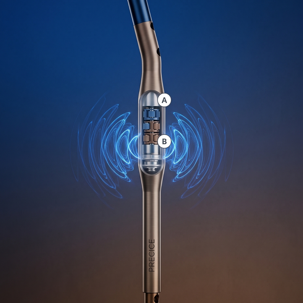
PRECICE – A Revolution in Limb Lengthening Without External Stabilizers