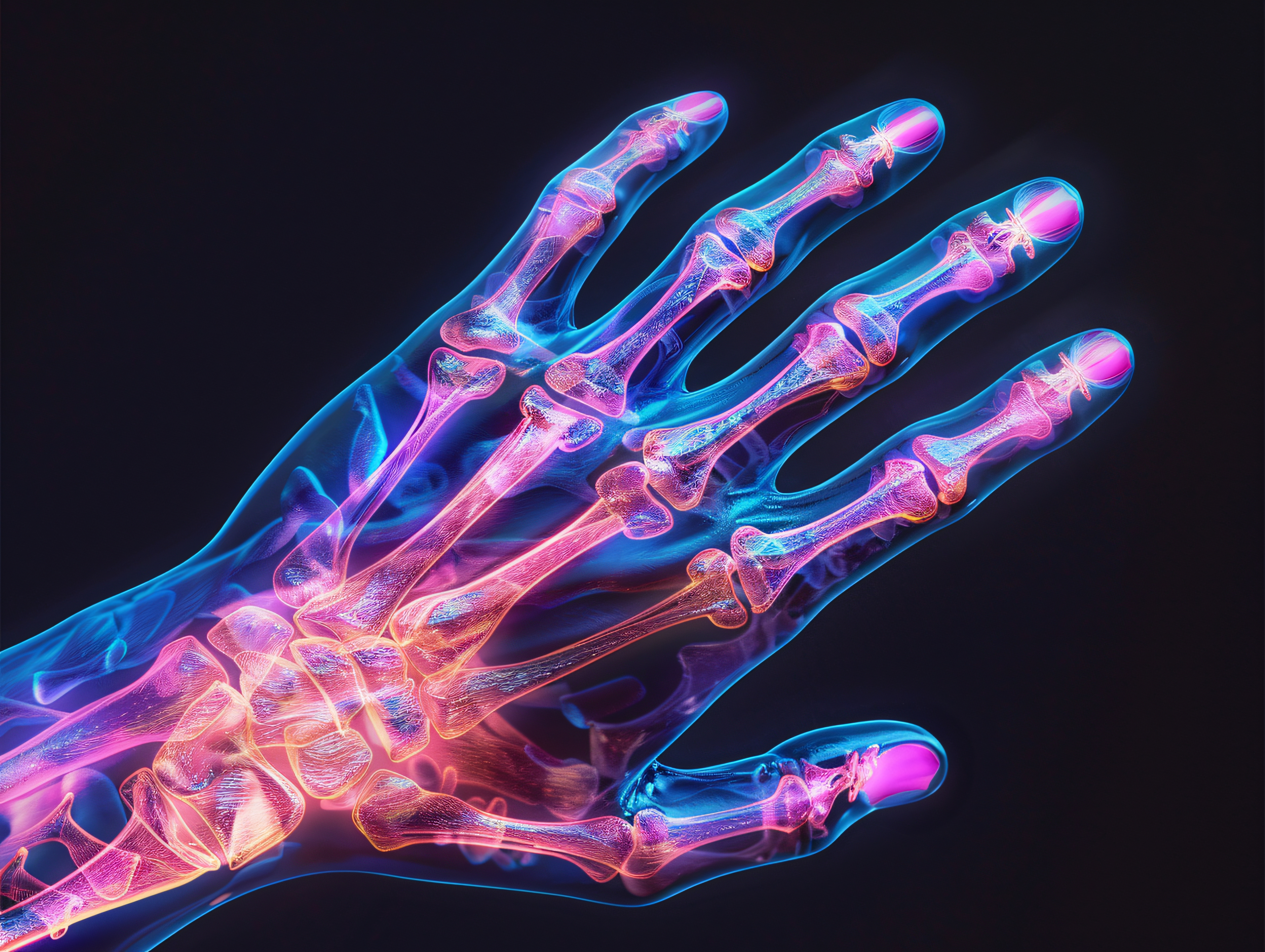
Hollow Foot – Symptoms, Causes, and Treatment Options
A high-arched foot (pes cavus) is a deformity in which the longitudinal arch of the foot is excessively arched. The mere presence of a high arch does not always indicate a medical condition—for some people, it is simply a variation in foot structure that does not cause any symptoms. The problem arises when the increased arch is associated with abnormal weight distribution, pain, ankle instability, toe deformity, or impaired gait. A pes cavus in a child requires special attention, as in some cases it may be the first symptom of a neuromuscular disorder.
At the Paley European Institute, the diagnosis of foot deformities takes into account the patient’s age, the cause of the problem, the extent of the deformity, and its impact on function. This allows us to determine whether conservative management will be sufficient or whether more advanced orthopedic treatment is necessary.
What is a hollow foot?
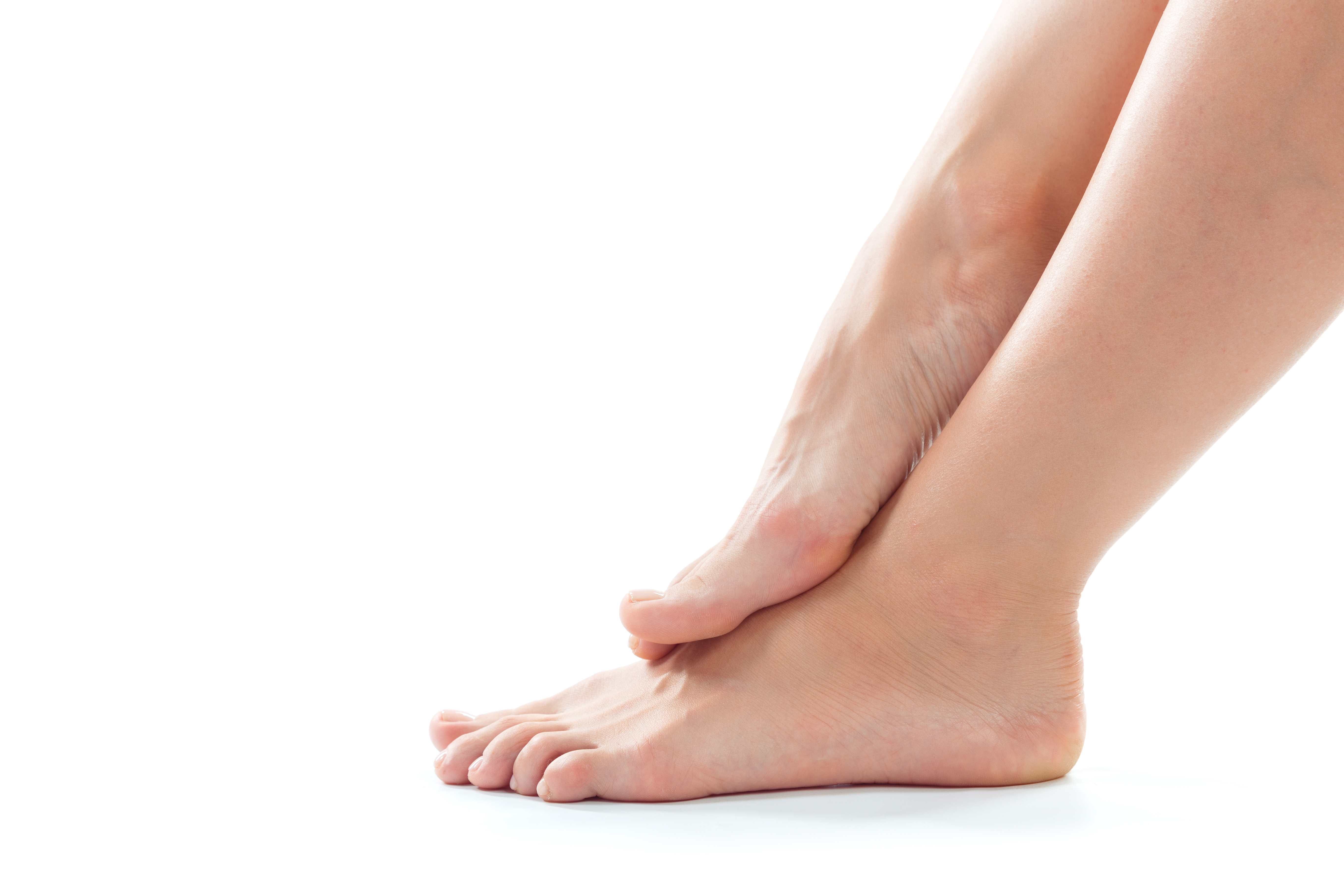
A hollow foot is characterized by excessive arching of the medial longitudinal arch. This deformity can affect one or both feet and vary in severity. In more complex cases, the high arch is accompanied by a varus heel, forefoot adduction, plantar flexion of the first metatarsal, and claw-like toe alignment. This configuration is often referred to as a cavovarus foot.
In a properly functioning foot, the load is distributed among its various parts during walking. With a hollow foot deformity, this mechanism may be disrupted. Excessive pressure on the forefoot or the lateral edge of the foot can lead to the formation of painful calluses, overuse of soft tissues, and an increased risk of ankle sprains.
Nor should a hollow foot be equated with all other foot deformities. For example, clubfoot is a distinct, multifaceted deformity that has its own diagnostic criteria and treatment methods.
What are the symptoms of hollow foot?
Symptoms depend on the severity of the deformity, its cause, and whether the foot remains flexible or becomes stiff over time. In some people, a hollow foot may not cause significant discomfort for a long time. In other cases, abnormal pressure distribution while walking leads to pain and functional problems.
Possible symptoms include:
- a distinctly arched foot,
- pain in the forefoot or heel,
- painful calluses on the sole of the foot,
- frequent sprains and a feeling of instability in the ankle joint,
- difficulties in choosing the right shoes,
- claw-like or hammer-like toe alignment,
- walking on the side of the foot,
- decreased tolerance for prolonged walking or running,
- gait abnormalities.
In children , progressive changes in the shape of the foot, frequent stumbling, a worsening gait, muscle weakness, or recurrent ankle sprains should also raise concerns. Such symptoms may indicate that the deformity is not merely a mechanical problem and requires further diagnostic evaluation.
Hollow Foot—What Are the Causes?
The causes of deformities vary. It is particularly important to rule out a neurological cause, especially when the deformity is bilateral, progressive, or occurs in a child without an obvious cause. According to the Pediatric Orthopaedic Society of North America, a diagnosis of idiopathic hollow foot should be made only after ruling out a possible neurological cause.
One of the most commonly associated causes is Charcot-Marie-Tooth disease (CMT), a hereditary peripheral neuropathy. Among other things, this condition causes weakness and an imbalance in the functioning of specific muscle groups, which can lead to the development of a hollow-knuckle deformity.
Other possible causes include:
- other neuromuscular disorders,
- spinal cord disorders, including syndromes associated with spinal cord entrapment,
- the consequences of certain congenital defects,
- residual effects of other foot deformities,
- consequences of injuries,
- muscle imbalance,
- idiopathic cases in which no clear cause can be identified.
Therefore, treating the foot alone does not always solve the problem. If the deformity is caused by a progressive neurological disease, it is necessary to take this into account when planning further treatment.
How is a hollow foot diagnosed?
Diagnosis begins with a thorough medical history and physical examination. The specialist assesses, among other things, the height of the arch, the position of the heel and forefoot, the range of motion in the joints, the presence of contractures, the condition of the skin, and how weight is distributed on the foot. Gait analysis is also an important part of the process.
In patients with a suspected neurological cause, it is also necessary to assess muscle strength, reflexes, sensation, and other symptoms that may indicate a disease of the nervous system. Information about similar problems in family members is also important.
One of the tests used to assess the flexibility of deformities is the Coleman test. It helps determine whether a hindfoot varus deformity is flexible—and depends primarily on the position of the forefoot—or whether it is rigid. This distinction can be important when planning treatment.
Depending on the situation, imaging tests may also be performed, primarily X-rays of the foot. If a neurological condition is suspected, the doctor may decide to expand the diagnostic workup to include a neurological consultation, electrophysiological tests, imaging of the spine, or other tests appropriate for the suspected cause.
Hollow Foot in Children – When Should You Seek Medical Advice?
A hollow foot in a child requires special attention, as its cause is not always apparent during the initial examination. The literature on children and adolescents emphasizes that, in cases of hollow-valgus foot, neurological disorders must be ruled out.
A consultation is particularly recommended when:
- the deformity develops or worsens with age,
- The problem affects both feet,
- The child often trips,
- there are recurrent ankle sprains,
- pain occurs when walking or during physical activity,
- claw-like fingers are visible,
- muscle weakness occurs,
- There was a history of neuromuscular diseases in the family.
In such situations, it is advisable to have your child examined by a specialist. A pediatric orthopedist will assess the nature of the deformity and determine whether further diagnostic testing is needed. If a neurological disorder is suspected, the course of action should also include an appropriate consultation.
However, this does not mean that every foot with a high arch is a sign of a medical condition. In children, there may also be an asymptomatic form of high arch, which is why a comprehensive evaluation of the patient is important, rather than focusing solely on the appearance of the foot.
How is a hollow foot treated?
The treatment approach depends primarily on the cause, the severity of the deformity, its flexibility, and the presence of pain and functional limitations. Not every hollow foot requires surgery. In cases of mild or stable deformities, the doctor may recommend observation and conservative management.
Non-surgical treatment may include:
- properly fitted shoes,
- custom orthotics or insoles,
- stretching exercises for contracted structures,
- strengthening weakened muscle groups,
- physical therapy aimed at improving balance and motor control,
- adjusting activity levels in cases of pain.
It is particularly important to customize orthotic devices. In cases of symptomatic pes cavus, a properly designed orthosis can help redistribute pressure and improve stability. A Cochrane review points to the potential benefits of using custom-made orthoses in alleviating pain associated with pes cavus, although the authors emphasize the limited quality of the available evidence.
When might surgery for a hollow foot be necessary?
Surgical treatment is primarily considered when the deformity is progressive, causes pain, leads to significant instability, or when conservative methods fail to restore satisfactory function. It is also important whether the deformity is flexible or fixed.
The surgery does not involve a single, one-size-fits-all procedure. The scope of the surgery is tailored to the specific deformity. Depending on the case, the following procedures, among others, may be used:
- releasing tight soft tissues, including the plantar fascia,
- stretching of selected muscle-tendon structures,
- tendon transfers to improve muscle balance,
- metatarsal osteotomies,
- metatarsal or tarsal osteotomies,
- correction of the calcaneus position,
- in cases of advanced, rigid deformities—more extensive reconstructive procedures.
In children, it is particularly important to consider growth potential and the cause of the deformity. In the case of a progressive condition, simply correcting the foot’s alignment may not be sufficient—it is also necessary to address the neuromuscular disorders that led to the deformity. The goal of treatment is to achieve a foot that is as pain-free, stable, and functional as possible, while preserving mobility whenever possible.
Treatment of Clubfoot at the Paley European Institute
At the Paley European Institute , clubfoot is evaluated in the context of the entire musculoskeletal system and the possible cause of the deformity. This is particularly important in children, in whom the changes may be related to neuromuscular disorders and may evolve as the patient grows.
The diagnostic process may include a clinical evaluation, gait analysis, imaging studies, and—when warranted—further diagnostic testing for neurological disorders. Based on the results, the treatment goal is determined and an appropriate course of action is selected.
Depending on the situation, treatment may include rehabilitation, physical therapy, orthotic devices, or surgery. This approach is consistent with the principle of a stepwise treatment plan—starting with the least invasive methods and progressing to surgical reconstruction when the deformity is severe, painful, or progressive.
For patients requiring surgical treatment, orthopedic surgery may include procedures aimed at both correcting bone deformities and restoring soft-tissue balance. The scope of the procedure always depends on the anatomy of the foot, its range of motion, the cause of the deformity, and the function of the entire limb.
A hollow foot should not be evaluated solely based on its appearance. If it causes pain, difficulty walking, instability, or gradually worsens, a thorough evaluation is warranted. It is particularly important in children to determine the cause as early as possible, as this allows for treatment tailored to their stage of development and helps reduce the risk of the deformity becoming permanent.
Hollow Foot in a Child – Consultation with Dr. Jacek Kapiński
At the Paley European Institute, we diagnose and treat foot deformities in children, tailoring our approach to the type of deformity, its severity, and its impact on the patient’s functioning. Orthopedic consultations for children are conducted by, among others, Dr. Jacek Kąpiński— a pediatric orthopedist specializing in congenital and acquired foot deformities as well as other orthopedic conditions in children and adolescents.
Dr. Jacek Kąpiński sees patients from birth through age 18. In his practice, he specializes in foot deformities, among other conditions, and has gained particular expertise in treating clubfoot using the Ponseti method. He is also a physician recommended by Ponseti International (we also encourage you to review the social media profiles of our pediatric orthopedist). If a child’s hollow foot is progressive, causes pain, walking difficulties, or frequent sprains, or is accompanied by muscle weakness, an orthopedic consultation can help assess the deformity and determine whether further diagnostic testing is needed.
Medical Sources
This study drew on, among other sources, current specialized materials and publications on the diagnosis and treatment of pes cavus:
- Pediatric Orthopaedic Society of North America – Cavus Foot
- American Orthopaedic Foot & Ankle Society – Cavus Foot Surgery
- EFORT Open Reviews – How to Manage Pes Cavus in Children and Adolescents?
- Cochrane – Interventions for the Prevention and Treatment of Pes Cavus
- Evaluation and Management of Cavus Foot in Adults – PMC
See other entries
How to Properly Care for a Stump After Amputation? A Practical Guide
Hollow Foot – Symptoms, Causes, and Treatment Options