Equinus foot
Equinus foot - the second most common congenital defect in children after hip dislocation. It can also be an acquired defect, secondary to external forces or neuromuscular disorders. It is a complex deformity of the foot, which is characterized by excessive hollowing of the longitudinal arch of the foot (the foot looks as if it is "broken" in half), adduction of the foot (pointing inward), heel splay (the foot rests on its outer edge) and horse heel bone alignment (the child looks as if he is standing on his toes all the time). Some muscles in the calf on the side of the foot with the defect atrophy. The etiology of the lesions is not fully understood. It is assumed that the clubfoot arises as a result of a combination of genetic and environmental factors. A normally developing foot turns into a limp-foot in the second trimester of pregnancy. Approximately 100,000 children are born with this defect per year, with twice the incidence in boys.
Typical equinus feet:
- Idiopathic - the cause of its origin is unknown; it is an isolated defect - there are no other defects or diseases besides it;
- syndromic - the cause of its formation is genetic mutations that cause birth defects; it often occurs in association with Pierre-Robin sequence, diastrophic dysplasia, arthrogryposis, etc;
- neurogenic - it occurs in conjunction with congenital defects on neurological grounds, such as cerebral palsy, spina bifida, spinal cord anchorage;
- teratogenic - its basis is disorders arising during pregnancy, for example, as a result of viral infections, taking drugs (amphetamines), certain medications, smoking, drinking alcohol or poisoning with toxins.
Atypical equinus feet - in addition to the standard features, they have a characteristic deep furrow over the heel and on the sole of the foot, hyper-extension of the big toe, very strong flexion of the sole of the foot and equine alignment. Types:
- atypical - occurs only in the idiopathic variety, can be diagnosed immediately after birth;
- complex - can involve all types of typical equinus deformities, and is the result of poor casting technique with the addition of complications while wearing the cast.
The treatment of equinus foot is a long-term process with multiple stages. Deformed feet differ in the degree of stiffness, muscle involvement, bony deformities and response to treatment. Therefore, the appropriate treatment method should be selected individually. Physical and radiological examinations are performed, which provide valuable knowledge about the severity of the defect and the need for surgical interventions.
The most popular method of treatment is the Ponseti method. It involves bringing the foot into natural alignment by modeling its shape with a cast (6-9 weeks), followed by wearing specially connected shoes (from several months to 5 years). It is also necessary to begin as soon as possible after birth exercises to increase the range of motion in the joint. The use of this method makes it possible to achieve a pain-free, well-functioning foot and removes restrictions on physical activity. The inability to use this method (for example, in neurogenic types of clubfoot, including arthrogryposis), the lack of progress during functional therapy, and the high severity of the defect are criteria for indications of surgery. Methods supporting the treatment of the clubfoot are neurophysiological physiotherapy using Prof. Vaclav Vojta's method of streamlining (stimulating muscle tension in a dynamic chain) and dynamic patching (increasing or decreasing muscle fiber tension with tape).
In about one-third of the cases, despite initially good correction of the defect, the patient again develops a talus and equine alignment. The main reason is premature termination of Denis-Brownea splint treatment. In this case, surgical intervention is required, involving various procedures, including lengthening of the Achilles tendon or posterior tibialis muscle.
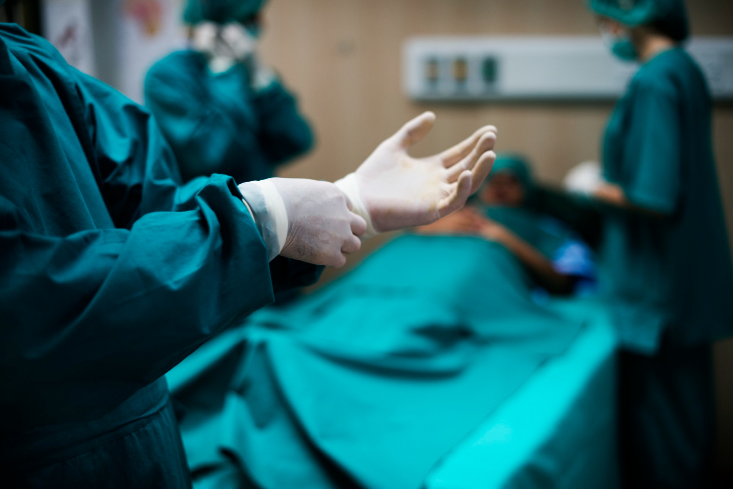
At Paley European Institute, we comprehensively treat foot deformities. We have top specialists who perform surgical operations for foot deformities, including recurrent clubfoot, provide physiotherapy, and supply patients with orthotic supplies.
See other entries
What is SEMLS surgery, and who needs it?
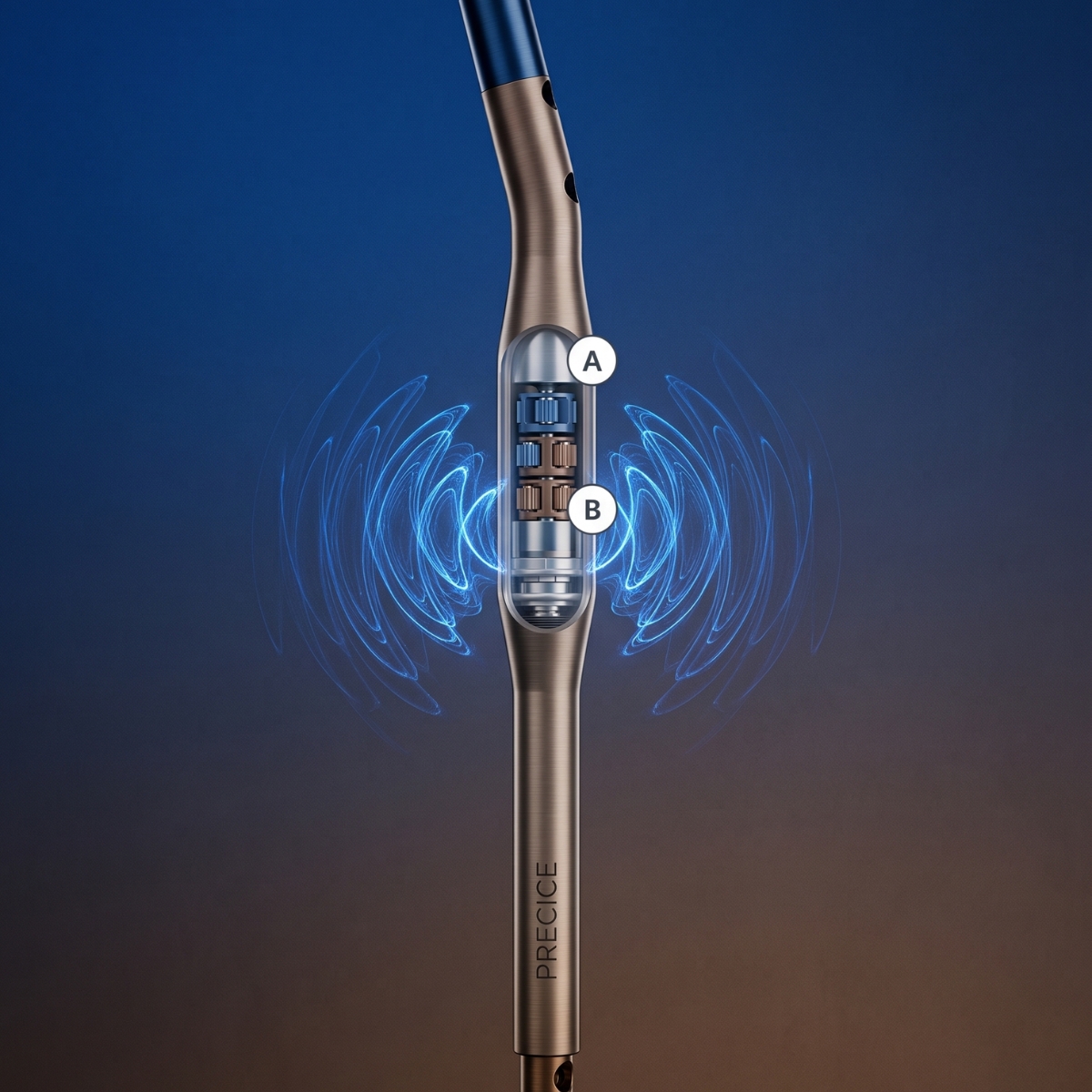
PRECICE – A Revolution in Limb Lengthening Without External Stabilizers