Hip Disorders in Infants and Children—Dysplasia, Hip Dislocation, Perthes Disease
At the Paley European Institute, we comprehensively diagnose and treat all types of hip disorders in children. We begin with thorough imaging studies and tailored conservative treatment, and when necessary, we use Dr. Dror Paley’s advanced surgical techniques. Early diagnosis and appropriate treatment are crucial for the proper development of the hip and the child’s mobility in adulthood.
What are hip disorders in children?
Hip disorders in children are a group of conditions in which the hip joint develops or functions abnormally, which can lead to instability, dislocation, or deformity. The hip joint is formed by the femoral head and the acetabulum. In infants, both structures are still largely cartilaginous and continue to develop during the first months and years of life. Therefore, a disorder detected early can usually be treated in a simple, noninvasive manner.
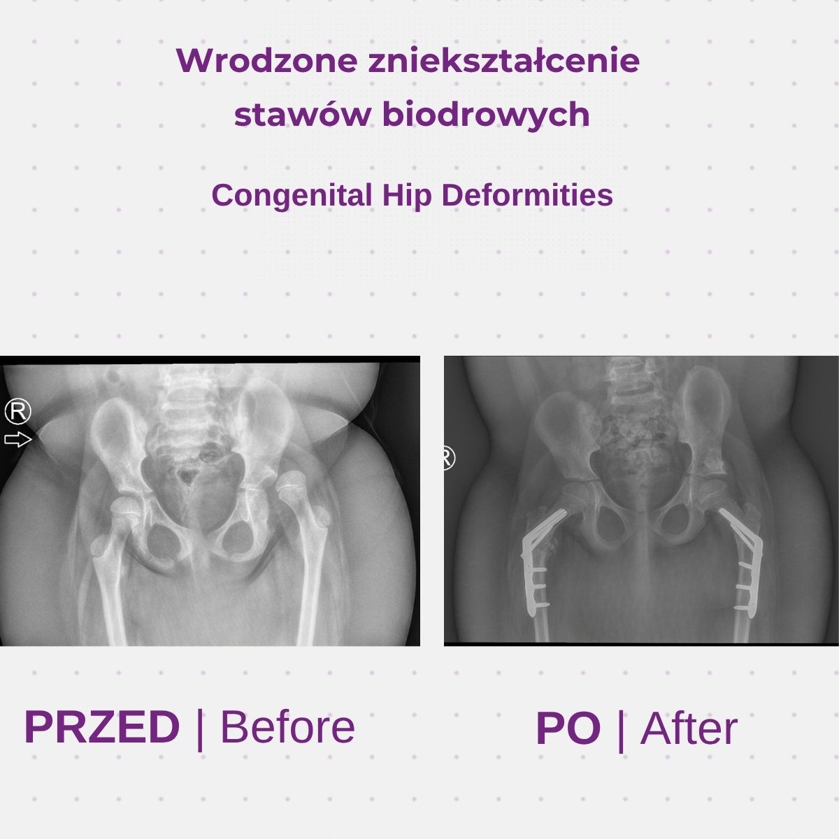
Disorders of the hip joint in infants and children
Symptoms
In infants, hip disorders often do not present with obvious symptoms. Parents should be concerned if they notice:
- limited or asymmetrical leg abduction
- asymmetry in skin folds on the thighs and buttocks
- one leg appearing shorter than the other
- in older children: limping, pain in the hip, groin, or knee
- getting tired quickly while walking
Reasons
The causes depend on the type of condition. Hip dysplasia and hip dislocation are most often associated with abnormal fetal positioning in the uterus. Perthes disease results from impaired blood flow to the femoral head. Some of these conditions are associated with congenital defects and neurological disorders.
Diagnostics
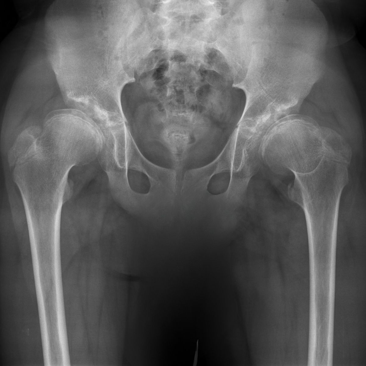
The basis for diagnosis is an orthopedic examination. In infants, a Graf hip ultrasound is performed, which in Poland is part of the newborn screening program. In older children, X-rays are used, and in complex cases, magnetic resonance imaging (MRI).
The Global Standard for Pediatric Hip Treatment at the Paley European Institute
Paley European Institute treats hip disorders in children using the methods of Dr. Dror Paley , one of the world's most experienced specialists in pediatric orthopedics, limb reconstruction and lengthening.
The Institute is the European center of the Paley Orthopedic & Spine Institute in West Palm Beach, Florida, which has one of the largest experiences in the US in the surgical treatment of hip dysplasia and dislocation.
’s Team of Pediatric Orthopedic Specialists
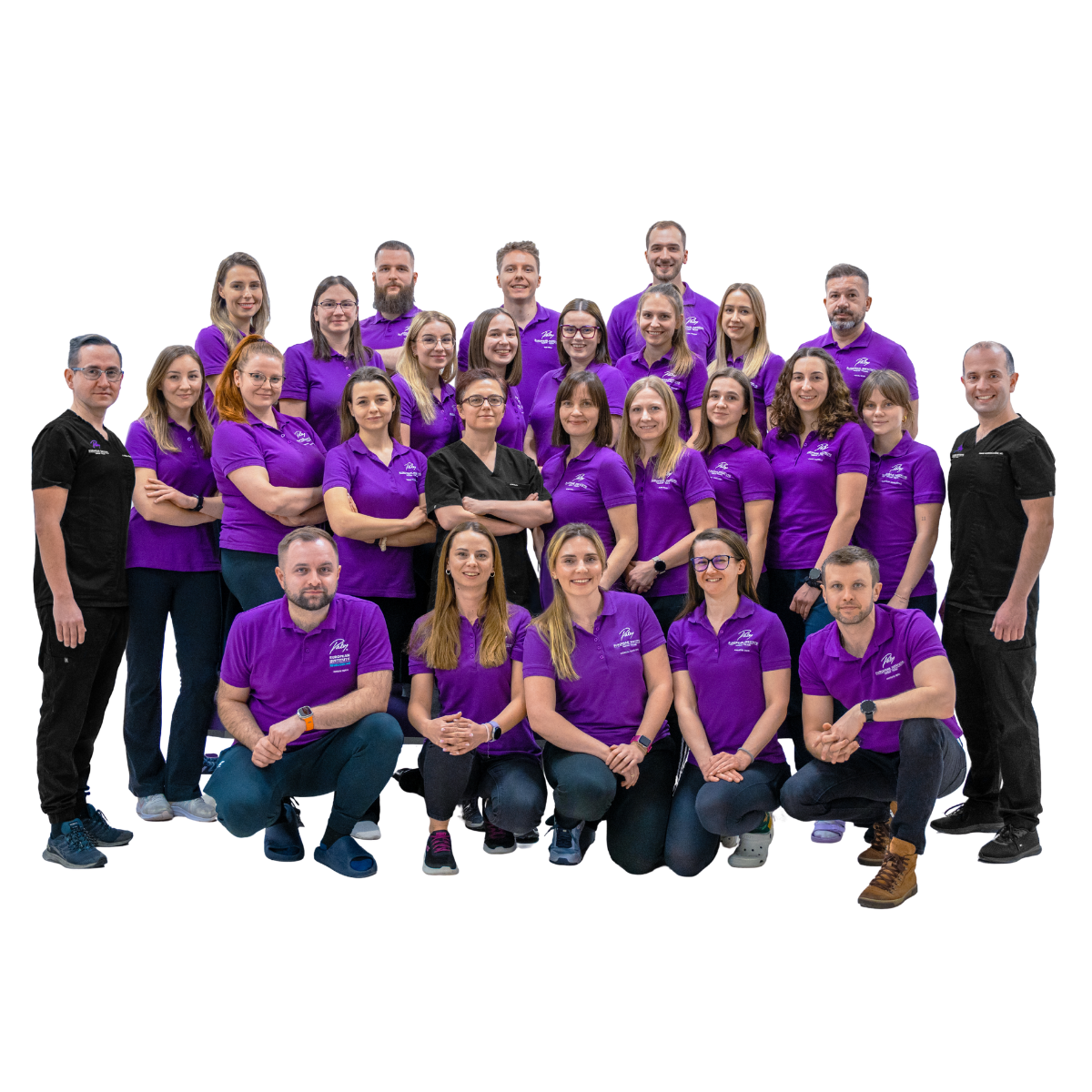
Patients are cared for by pediatric orthopedic specialists in collaboration with physical therapists and radiologists. The team supports the child from the first ultrasound, through treatment, until the hip joint has fully developed.
Simultaneous Bilateral Hip Reconstruction
At the Paley European Institute, children with bilateral dysplasia or dislocation can undergo simultaneous reconstruction of both hips in a single surgery. The child undergoes one anesthesia, one immobilization, and one rehabilitation period instead of two.
When should you see a pediatric orthopedist if you suspect hip dysplasia?
You should take your child to a pediatric orthopedist as early as possible—preferably within the first few weeks of life—and at any age as soon as you notice any concerning symptoms. The earlier treatment begins, the simpler and more effective it is.
A consultation is particularly recommended when:
An ultrasound of the hips revealed abnormalities
The baby was born in a breech position
There was a history of hip dysplasia in the family
The baby's legs do not spread out symmetrically, or the folds on the thighs are uneven
The child limps, walks with a waddling gait, or complains of hip or knee pain
A child between the ages of 4 and 8 has started limping for no apparent reason
Previous conservative treatment was ineffective

How do we treat dysplasia and other hip disorders in children?
Treatment is tailored individually to the type of disorder, its severity, and the child’s age. It includes diagnosis, conservative treatment, and, if necessary, surgical treatment, rehabilitation, and regular monitoring of hip joint development.
Diagnostics
orthopedic examination
imaging diagnostics: ultrasound, X-ray, MRI
evaluation of newborn screening results
Conservative treatment
Pavlik harness, which keeps the legs in a flexed and abducted position; used during the first months of life, typically for a few weeks to a few months under ultrasound monitoring
abduction braces
extensive diaper use for mild forms
In older infants: closed reduction under anesthesia and a hip cast, usually for about 6 weeks
physical therapy
Surgical treatment
open reduction, i.e., placing the head of the femur into the acetabulum
pelvic osteotomy, which improves the coverage of the femoral head
by the acetabulum
femoral osteotomy, when necessary to achieve proper joint alignment
a slight shortening of the femur in cases of severe dislocation; the difference in length corrects itself spontaneously during growth
reconstruction of the oblique ligament, which stabilizes the femoral head within the joint
simultaneous bilateral hip reconstruction
Postoperative Care
a hip cast applied while the patient is still in the operating room
hospitalization, usually for 3–4 days
Follow-up examination of the wound after about a week and, if possible, replacement of the cast with a removable brace
An X-ray taken after about 6 weeks to assess the treatment outcome
Rehabilitation and Regular Monitoring
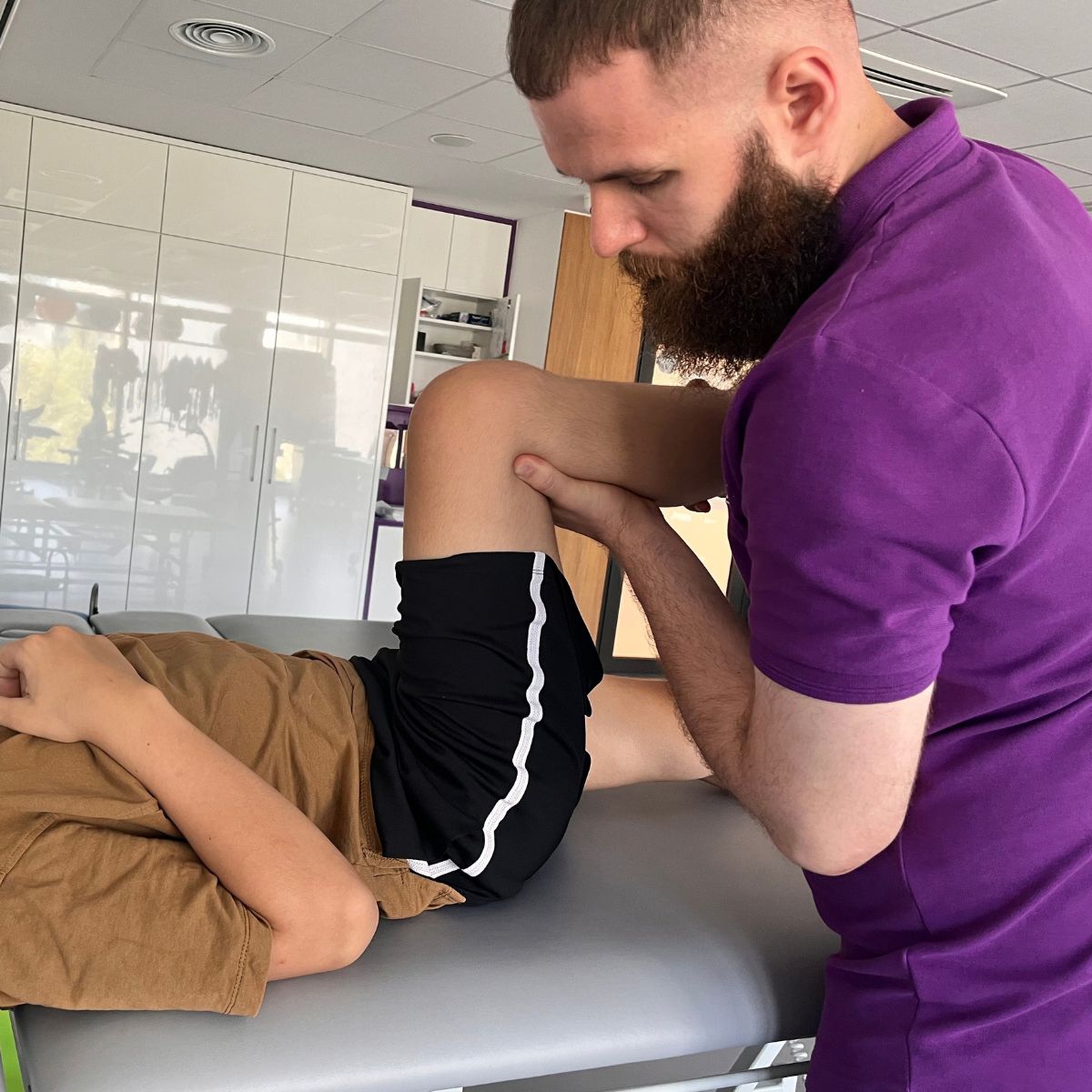
Physical therapy to strengthen the hip and leg muscles and restore range of motion
Regular checkups on hip joint development until growth is complete
What challenges do children with hip disorders and their parents face?
The biggest challenge is that dysplasia in infants often has no symptoms, and if left untreated, it leads to permanent changes in the joint.
Late diagnosis. When dysplasia is detected only after the dog has started walking, conservative treatment is often no longer sufficient.
Limping and pain. A misaligned hip causes limping, rapid fatigue, and pain.
Difference in limb length. A dislocated hip makes the leg functionally shorter.
Early-onset osteoarthritis. Untreated or inadequately treated dysplasia can lead to joint degeneration even in young adults and the need for joint replacement surgery.
A long recovery. Braces, casts, and physical therapy require patience and the commitment of the whole family.
Thanks to early diagnosis and well-planned treatment, most children develop normally and can walk, run, and play sports without any limitations.
Hip dysplasia
Hip dysplasia is the abnormal development of the hip socket, which can lead to hip instability or dislocation. The socket is shallow and does not properly enclose the femoral head, causing the joint to become loose and not develop as it should.
Hip dysplasia occurs in approximately 1–2% of newborns, more often in girls, and in approximately 20% of cases affects both hips. If detected in the first weeks of life, it can usually be treated non-invasively, for example, with a Pavlik harness.
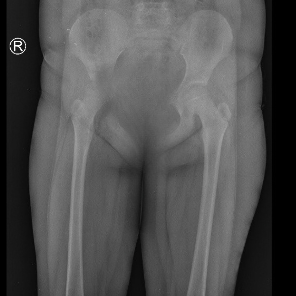
Congenital dislocation of the hip joint
Congenital hip dislocation is a condition in which the head of the femur is not properly seated in the socket at birth. It is currently considered the most severe form of developmental dysplasia of the hip (DDH) and affects approximately 1 in 1,000 births.
A dislocated hip causes limited movement and a shortened leg, and in children just beginning to walk, a limp. Treatment depends on age: braces or a cast are used for infants, while older children usually require surgery.
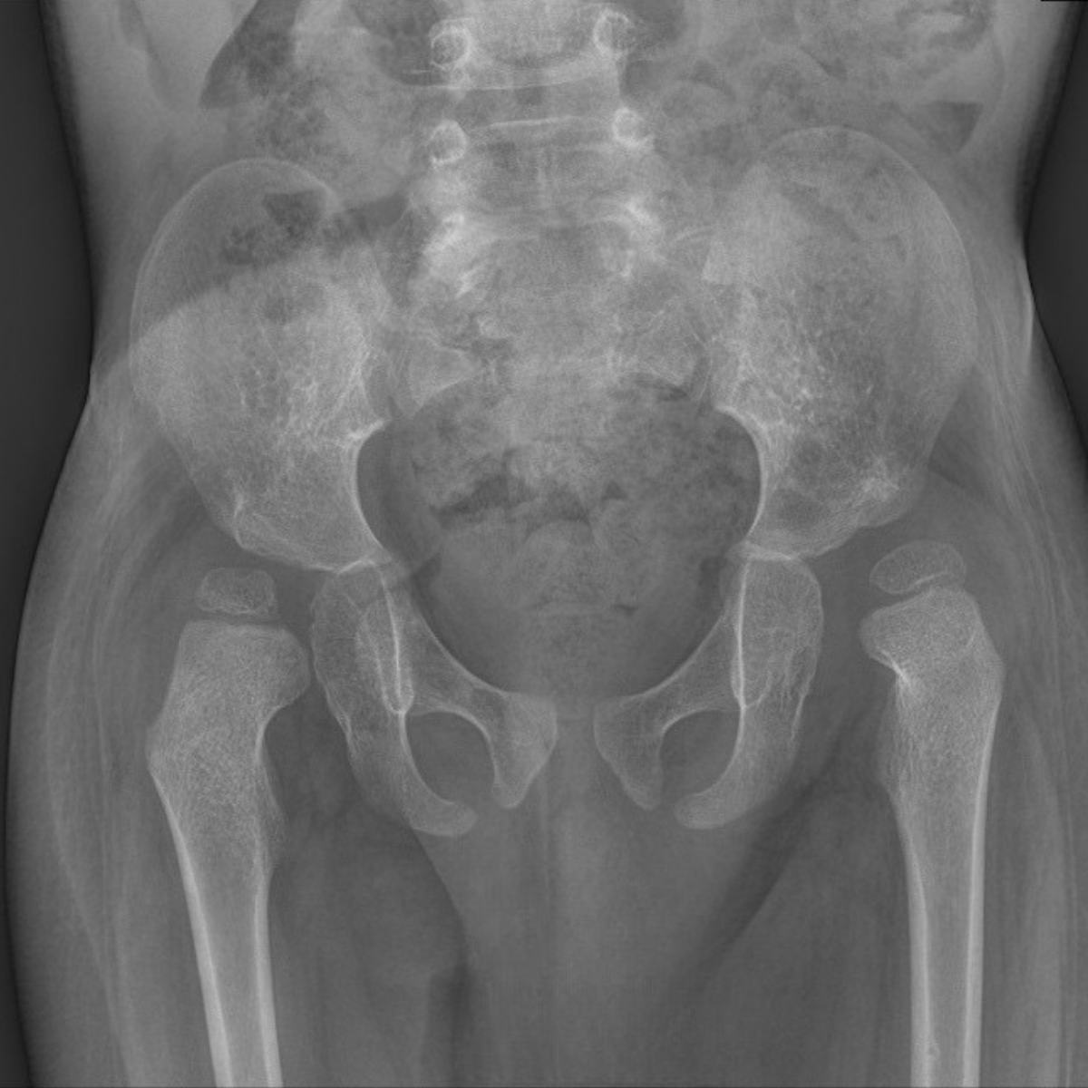
Legg-Calvé-Perthes disease
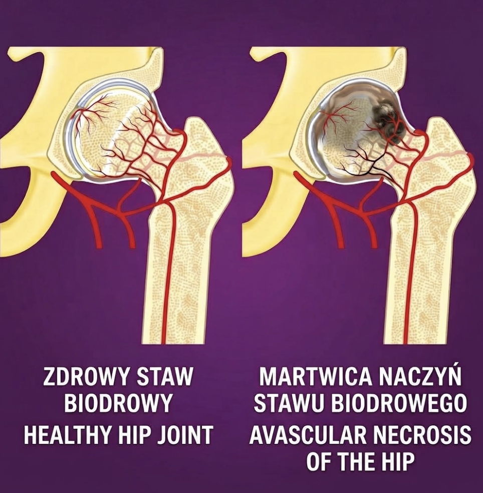
Legg-Calvé-Perthes disease is a temporary disruption of blood supply to the femoral head, leading to its necrosis and deformity. It most commonly occurs in children aged 4–8 years, and is several times more common in boys.
The first symptom is usually a limp and pain in the hip or knee. The bone rebuilds over time, and the goal of treatment is to ensure that the femoral head maintains its spherical shape and fits properly in the acetabulum.
Our Experts: Meet the Paley European Institute team today

Family-Centered Care: treatment in which the family is part of the care team
Family-Centered Care (FCC, family-centered care) is the treatment model used at the Paley European Institute. The family of a child with a congenital limb defect is not merely an observer but a full-fledged member of the treatment team. Treatment for congenital limb defects often lasts for months or years and involves several surgeries, limb lengthening, and months of rehabilitation. Therefore, parents who understand the treatment plan and feel heard have a real impact on its outcomes.
What does family-centered care look like in practice?
Parents do not wait in the hallway—while their child is undergoing surgery, the Hospital Services Coordinator and a physician’s assistant keep parents updated on the progress of the procedure via WhatsApp. During this time, a psychologist stays by the parent’s side to support them through this most difficult hour.
One treatment plan instead of multiple visits—during the consultation, specialists from various fields meet with the child in one place and develop a comprehensive treatment plan. The conversation begins not with the question “What hurts?”, but with what the child likes to do and what the family’s daily life is like.
The same physical therapist before, during, and after surgery—the therapist present in the operating room—conducts rehabilitation starting on the first day of recovery. The child exercises with someone they already know, which reduces anxiety and facilitates a return to physical fitness. This is particularly important during limb lengthening, when systematic physical therapy is crucial for maintaining joint mobility.
Comprehensive care in one place—X-rays, blood tests, an orthotist, a psychologist, a dietitian, a speech therapist, and certificates for schools and government offices—are all available without unnecessary referrals or having to search for other facilities. Surgeries are performed at our partner hospital, Medicover, where the Paley European Institute team cares for the patient from admission through discharge.
Support for families from other cities and countries—families traveling here for medical treatment receive assistance with arranging transportation, accommodations, and all the logistics of their stay.
Parents' Council—parents who have themselves undergone treatment at the Paley European Institute—regularly test and evaluate new approaches, helping to shape the model of care.
Why does family involvement improve treatment outcomes?
Parents who understand the treatment plan are better able to follow rehabilitation recommendations at home and recognize signs that require contacting a doctor more quickly. Thanks to preparing the family for home care, children who undergo surgery at the Paley European Institute stay in the hospital for an average of only 2 days. A shorter hospital stay and less stress lead to a more peaceful recovery and a better quality of life for the entire family.

Related articles
Read the blog posts on this topic

The hip joint - diagnosis and therapy
Ultrasound of the Hips in Young Children
Invited for infant hip ultrasound
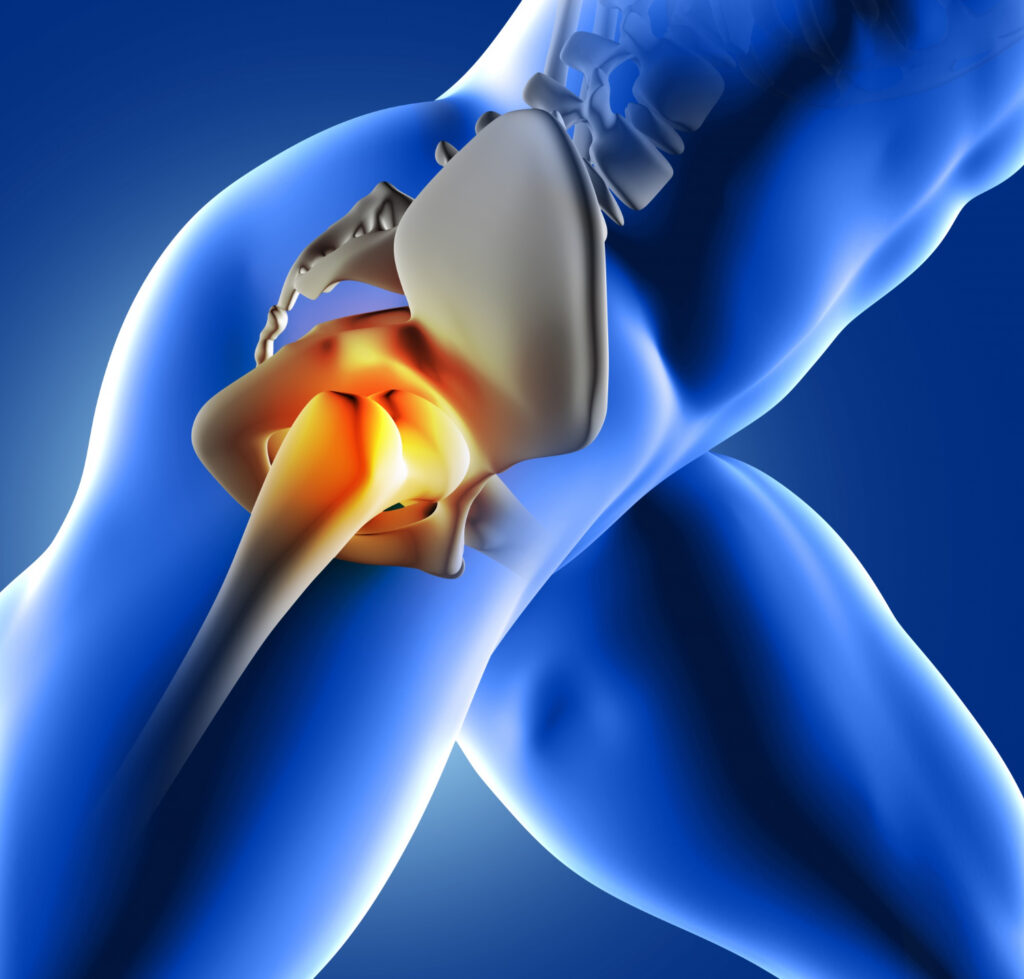
Hip Osteoarthritis – Symptoms, Management, and Treatment
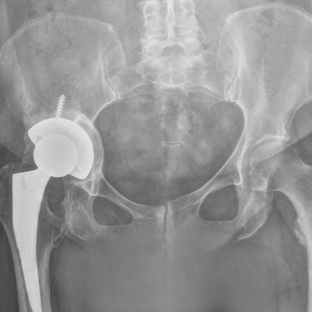
Developmental dysplasia of the hip joint
Your most frequently asked questions
That is, What you as a parent should know before making a decision.
This is an abnormal development of the hip socket that can lead to instability, subluxation, or dislocation of the hip. It affects about 1–2% of newborns, with girls being more commonly affected.
The limited range of motion in the legs, asymmetry in the thigh creases, and one leg that appears shorter are cause for concern. The diagnosis is confirmed by an orthopedic examination and a hip ultrasound.
The first ultrasound is performed as part of routine screening during the first weeks of life. The orthopedist determines the schedule for subsequent examinations.
Usually anywhere from a few weeks to a few months. The doctor determines the duration of treatment based on follow-up ultrasound exams.
Mild abnormalities in a newborn may resolve on their own, but they require ultrasound monitoring. Established dysplasia should not be left untreated, as it can lead to dislocation and joint degeneration.
When conservative treatment is ineffective or the dysplasia is diagnosed late, surgery is usually performed around the age of 2.
The femoral head is positioned within the acetabulum (open reduction), and an osteotomy of the pelvis—and sometimes the femur—improves the joint’s coverage and alignment. After surgery, the child wears a cast or brace and then undergoes physical therapy.
Meet Our Patients