Ultrasound of the Hips in Young Children
What is a hip ultrasound in children?
In the first months of life, every infant undergoes numerous screening tests aimed at detecting abnormalities early in life. Thanks to them, it is possible to quickly identify the problem and undertake treatment and rehabilitation. One of the recommended tests that should be performed is ultrasound (USG) of the hip joints in infants. They are performed to check the structure of the hip joint. The test can detect congenital hip dysplasia and assess the severity of the abnormality. This is one of the most common congenital abnormalities of the musculoskeletal system, which prevents the child from walking properly and consequently leads to impaired function of the entire musculoskeletal system.
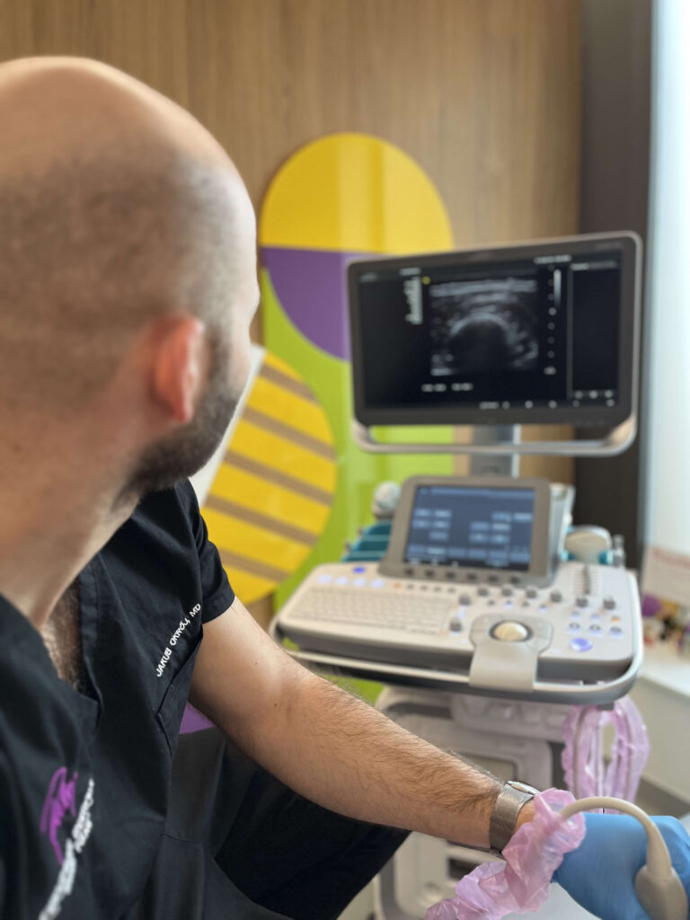
Infant hip ultrasound is a non-invasive imaging method that uses ultrasound to verify the condition of the hip joint cavity and surrounding soft tissues. It also provides information about the position of the femoral head relative to the acetabulum. During ultrasound of the hips in an infant, an ultrasound machine is used, which consists of a head and a monitor. It is on it that the doctor sees the image produced by the ultrasound waves.

Course of ultrasound of the hip joint in children
How does the ultrasound of the hip joints in children proceed? First of all, it should be noted that this is a painless and quick examination. It lasts about 10-15 minutes. The ultrasound of the hip joint in children takes place in a darkened room, so that the doctor can better see the image on the monitor. The specialist lubricates the skin around the hip joint with a special gel, over which he then moves the head of the device. This allows the ultrasound waves to penetrate deep into the tissues and then bounce off internal structures. The result of this process is an image of the joint seen in real time. The device also allows the taking of an image.
The next step is to evaluate the hip joint on the Graf scale. The doctor may find a normally developed hip (types Ia and Ib), an immature hip (including IIa), hip dysplasia (type IIc), dysplastic hip with femoral head decentration (type d), subluxated hip (types IIIa and IIIb) or dislocated hip (type IV). Importantly, the test results are obtained immediately after the ultrasound.
Indications for infant hip ultrasound
Ultrasound of the hip joints in children is performed by a pediatric orthopedist. It is performed as part of standard preventive examinations. Importantly, the examination should be carried out when the child has noticed worrying symptoms, such as:
- - Differences in appearance and leg length,
- - Limitations and problems with visiting (Barlow symptoms),
- - leg movement problems,
- - Joint skipping, or Ortolani's sign.
In addition, the test should be performed when a genetic predisposition to hip dysplasia has been identified, and when the baby was born from a buttock position. Most often, hip ultrasound in infants is performed between 6 and 12 weeks of age.
How do you prepare your child for the test?
The ultrasound of the hip joint does not require special preparations. The only thing that should be taken care of is the child's loose clothing, which can be easily removed and put on. A contraindication to the examination is an unhealed wound on the skin in the area where the examination will be conducted.
Why is it a good idea to perform hip ultrasounds on children?
Hip ultrasound can detect hip dysplasia
, which if left untreated has serious consequences. As a result, the child has problems with proper movement. After all, the hip joint has a huge impact on motor development and learning to walk. The earlier hip dysplasia is diagnosed, the greater the chance of a complete cure of the defect. Early introduction of treatment and physiotherapy will improve the condition of the joints. Therefore, hip ultrasound in children is an extremely important examination.
Diagnosed hip dysplasia at an early stage, and therefore quick conservative treatment, can cure an abnormally developed hip joint in 95% without surgical intervention.
Has your child been diagnosed with hip dysplasia? Get in touch with us! At Paley European Institute, you will receive professional help in the field of pediatric orthopedics.
See other entries

What is SEMLS surgery, and who needs it?
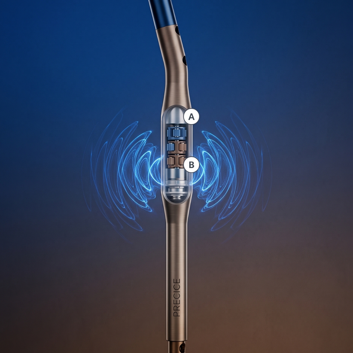
PRECICE – A Revolution in Limb Lengthening Without External Stabilizers