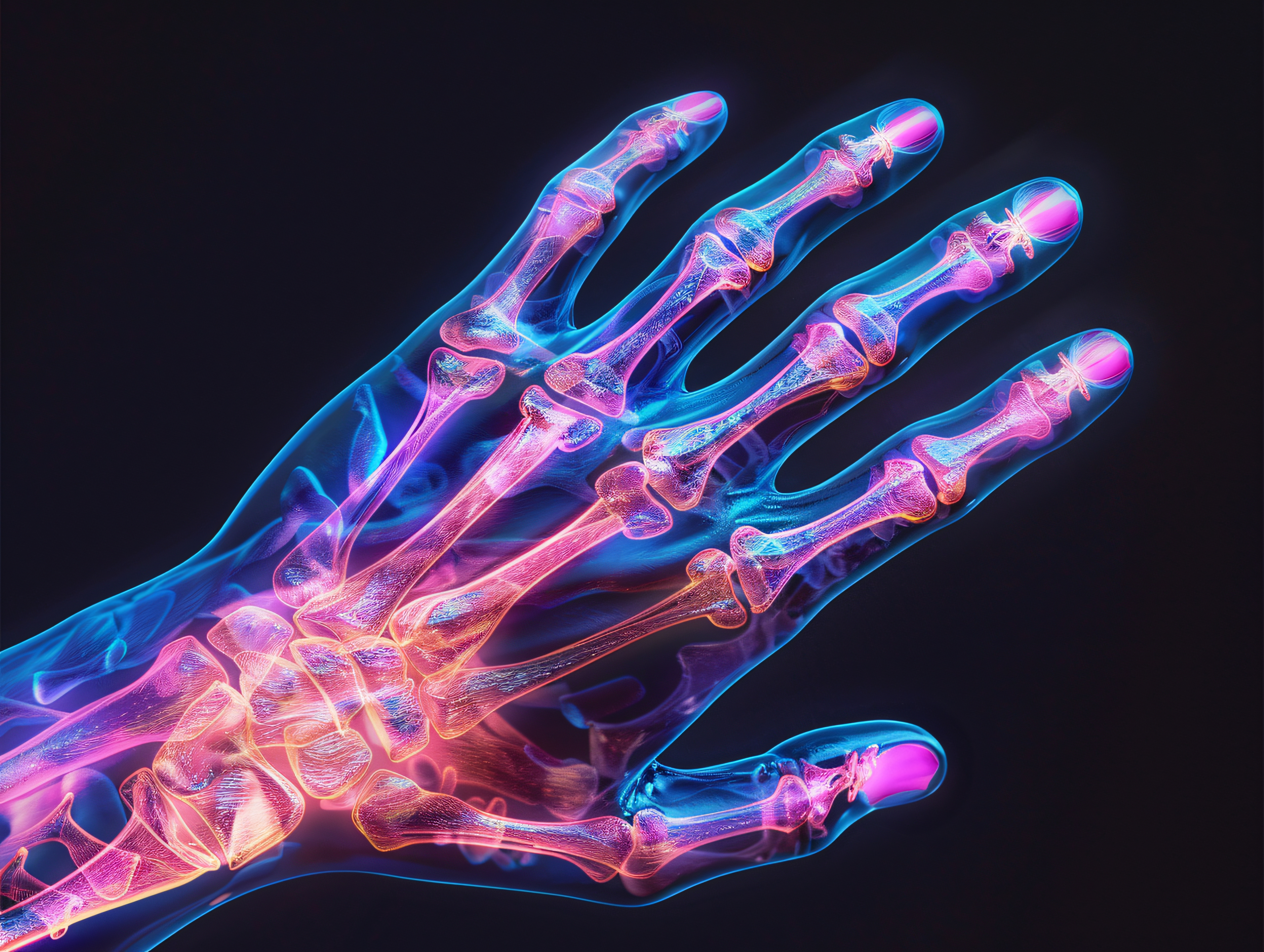
Dysplasias of the skeletal system (osteochondrodysplasias)
Osteochondrodysplasia is a disease characterized by an abnormal structure of the skeletal system. It results from abnormal growth, development and differentiation of bones and cartilage. Osteochondrodysplasias have a genetic basis and occur in 1 in 5,000 live births. Osteochondrodysplasia manifests as low height in childhood and disproportions in the body (except for dysplasias associated with a disorder of bone mineralization, where body proportions may be normal). The severity of symptoms can range from early arthritis in a person with average height to severe growth disorders causing prenatal death. Osteochondrodysplasia, depending on the cause, can be characterized by various dysmorphic features, such as a collapsed nasal root, hypoplasia of the midface, large-headedness, cleft palate or gray-colored irises. Bone dysplasia in children can cause significant limitations in daily functioning and even death.
In people with skeletal dysplasia associated with a bone mineralization disorder, body proportions may be normal. In contrast, disproportions may be seen in others.
Disproportionate short stature, resulting from osteoskeletal dysplasia, is divided into:
- The type with a short torso;
- The type with short limbs, in which we may have to deal with:
- rhizomelia, or shortening of the proximal parts (humerus and femur)
- mesomelia, or shortening of the middle parts (radius, elbow, tibia, fibula),
- acromelia, or shortening of the distal parts (hand, foot).
When discussing what dysplasia is, it is worth looking at the accompanying problems of osteochondrodysplasia, disorders stand out:
- respiratory system,
- cardiovascular system,
- excretory system,
- neuronal system,
- vision and hearing functions,
- psychological.
Osteochondrodysplasia - diagnosis and treatment
Dysplasia, even non-lethal dysplasia, can be detected as early as during an ultrasound performed during pregnancy. A precise history taking and detailed physical, radiological and genetic examinations are necessary for a correct diagnosis. Identification of the gene and the origin of skeletal dysplasia are essential to select treatment accordingly.
Most bone dysplasias have their own distinctive radiological features. In growing bones and cartilage in different types of osteochondrodysplasia are observed:
- In epiphyseal dysplasia - absent, small or irregular ossification of the bone epiphyses,
- In epiphyseal dysplasia - irregular, dilated appendages,
- In dysplasia involving the bone shafts - widening, sclerotization of the bone, thickening of the cortical layer of the bone, narrowing or widening of the marrow cavity.
Osteochondrodysplasia can be treated. The process is carried out with the cooperation of many specialists and should focus on minimizing complications, but also on aiming for the highest possible final height through the use of growth hormone therapy. It is extremely important to make a prompt and correct diagnosis. This allows timely introduction of treatment and prevention of functional deterioration.
Pediatric orthopedics focuses on specific features of dysplasia and includes:
- Prevention of bone deformities,
- joint stabilization,
- fracture prevention,
- Length compensation of the lower limbs,
- Prosthetics for joints affected by inflammation,
- decompression,
- correction of curvatures,
- Stabilization of the spine to prevent deformity and neurological damage.
It is also important to keep in mind possible breathing problems caused by a small chest circumference. In this case, it is necessary to introduce its support. It is also important to have regular hearing and vision check-ups, and to alleviate any pain that occurs. At Paley European Institute, we provide treatment, physiotherapy and orthotic support for patients with diastrophic dysplasia and growth dysphoria.
See other entries

How to Properly Care for a Stump After Amputation? A Practical Guide
Hollow Foot – Symptoms, Causes, and Treatment Options