Hypoplasia of the thumb
INFORMATION
A missing thumb or "thumb aplasia" is a complete absence of the thumb. Some patients may have a hypoplastic thumb or "floating thumb." In these patients, their thumb lacks the metacarpal bone and associated muscles and tendons. The thumb is attached only through the skin.
Congenital hypoplasia of the thumb is often associated with partial or complete absence of the radius bone. It occurs in 1:100,000 births with bilateral involvement (both hands) in 60% of patients. When one side is involved, the right hand is more common than the left.
Complete absence of a thumb can be an individual trait, but is often (>80%) associated with congenital abnormalities, such as:
The presence of the opposite finger is needed to manipulate everyday objects. Children born with this condition adapt quite well with few limitations. The decision to undergo surgical intervention therefore rests with the parents. Surgical treatment for complete absence of the thumb requires policyzation.
TREATMENT STRATEGY
Treatment of thumb aplasia and hypoplasia depends on the condition and nature of all the lesions associated with the
associated with congenital defects. In the case of aplasia or hypoplasia, the recommended method of treatment is policy surgery, which reconstructs the index finger into a thumb.
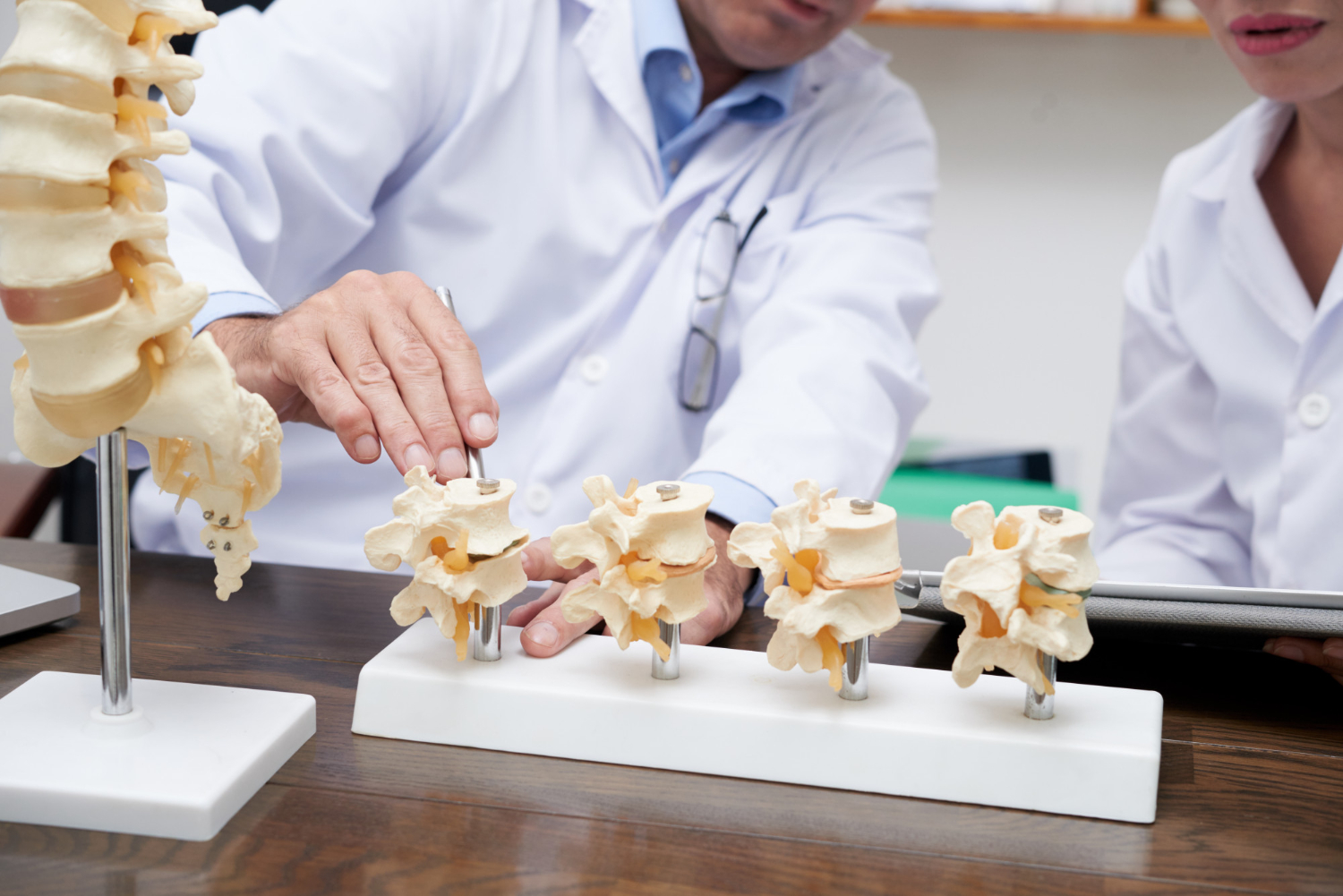
The classification system was developed in 1937 by Muller and improved by Blauth, Buck-Gramacko and Manske. The classification system includes 5 types:
Type 1
Type 2
and tendon transfer.
Type 3A
and tendon transfer.
Type 3B
Type 4
Type 5
The initial diagnosis includes an evaluation of the patient's hand, including tendon and skin abnormalities, joint stability and space tightness. This allows Dr. Paley or Dr. Robbinson to recommend an appropriate treatment plan. Evaluation of associated abnormalities is also essential during the initial consultation and may include cardiac, renal and abdominal ultrasound. To rule out Fanconi anemia, a blood smear and complete blood count may be recommended, as well as a chromosome provocation test.
In cases of thumb aplasia or hypoplasia, the recommended treatment strategy is policyz surgery.
POLICYZATION OPERATION
Dr. Paley treats a missing or hypoplastic thumb with a policyzation (surgical construction) of the index finger. Dr. Paley surgically moves the index finger to the thumb position. The metacarpal index finger (bone) is cut, and the finger is rotated and moved to the base of the hand in the normal thumb position.
It is important to preserve blood circulation and thumb sensation. Dr. Paley finds all veins, arteries and nerves, making sure that repositioning does not affect them. To restore thumb movement, muscles are moved to new locations. The thumb is usually rotated 135 degrees relative to the fingers. It is important to put it in the correct position so that the thumb, not the finger, works. The length of the thumb should be adjusted so that the tip of the thumb is at the level of the PIP joint. Too often, this operation is performed incorrectly when the thumb is improperly rotated, too long or with too much pressure. Dr. Paley is very careful when rotating the thumb to its most functional position and adjusting the length appropriately as a new finger. It is adjusted so that the thumb cannot straighten. This gives the thumb maximum stability and mobility and the two main functions of the thumb: pinching and opposition.
The combination of these steps gives the thumb an excellent cosmetic appearance. When policyzation or ulnarization is required, ulnarization is first performed a few months later either by policyzation or at the time of pin removal. In some cases, if the thumb muscles are not sufficient, Dr. Paley transfers muscles from other parts of the hand to improve movement.
ULNARIZATION (ULNARYZATION)
One of the main goals of Ulnarization surgery is to correct poor grip strength in people with RCH. Poor grip strength results from:
To understand this, try bending your wrist down, palm side down, and then try clenching your fist. You will find that it is much more difficult to clench your fist than with your wrist in a neutral position. For the repair, Dr. Paley relocates the ulnar flexor tendon (FCU), which makes the grip strength stronger.
In the Ulnarization technique, Dr. Paley turns the head of the elbow bone into a fulcrum. This makes recurrence impossible because the elbow physically blocks the wrist from returning to its deformed position. The FCU is transmitted from the palmar side to the dorsal side of the wrist. The FCU is a very strong tendon, and the transferred one becomes the new wrist extensor, allowing for increased grip strength and range of motion of the finger.
First, Dr. Paley makes an incision on the palmar side of the wrist to minimize soft tissue complications. The wrist is carefully cut out and the pea bone and FCU are released. If a radial fibular origin is present, it is again excised. To relocate the wrist, it must be disconnected from the ulna bone during a process called palmar capsulotomy. The head of the ulnar bone is completely detached from the wrist, but the muscles and blood vessels are preserved and protected. The head of the ulnar bone rests in the pocket of the radial bone, and the wrist is moved to the ulnar side. A pin is then inserted through the ulna and the cartilage of the wrist to keep everything in proper alignment. Then the FCU is moved and reattached to the cartilage of the fourth metacarpal bone. Internal K-wires are used to maintain the new alignment. The final step is to free the ulnar bone from the carpal joint. Dr. Paley usually uses an external stabilizer, but has now developed a new method using internal fixation.
POST-OPERATIVE PROTOCOL
After the procedure, the patient has a permanently elevated upper limb and is under constant neurovascular monitoring performed by staff every four hours. Pain and muscle spasms are constantly controlled. Expansion of the external stabilizer begins on the first day after surgery. Expansion is done with one to three screw rotations 3 times a day, for a total of 0.8 mm per day. Lengthening continues until the proximal end of the wrist is at the level of the apex of the distal epicondyle. Physiotherapy begins from the first day after surgery and includes exercises for elbow and finger mobility.
The external stabilizer is maintained for about three months, at which time it is removed in a smaller outpatient procedure. A wrist splint is constructed, which should be worn between therapy sessions at all times, for two months, and then only at night. Therapy continues after the splint is removed and should focus on achieving maximum range of motion in the wrist, elbow and fingers.
The Ulnarization technique makes the wrist joint completely mobile, with no relapse or growth arrest. These are remarkable results for Radial Club Hand. In conclusion, Ulnarization is a safe surgical technique that has proven:
See other entries

The Paley European Institute Wins Two Awards in the “Patient Trust Leader 2026” Competition

Bowlegs in Adults – Causes, Symptoms, and Effective Treatments