Juvenile sclerosis of the femoral head - SCFE
One of the most common diseases of the hip joint in childhood is slipped capital femoral epiphysis (SCFE). It involves the separation of a portion of the femoral head at the level of the growth cartilage, which is usually caused by circulation disorders in the cartilage area or trauma to the hip joint.
The incidence of juvenile femoral head desquamation is 2 : 100,000 live births and affects children aged 6-18 (most commonly 10-16). In most cases, the causes of SCFE are unknown. However, some authors identify risk factors and comorbidities. Among them are: poor eating habits, low physical activity, obesity, as well as hormonal disorders or genetic syndromes.
Hip pain in children, is it the only symptom of femoral head desquamation?
Juvenile femoral head desquamation mainly affects people in adolescence, when there is rapid body growth. Patients initially experience mild pain in the hip area, which may radiate to the thigh or knee. There is often a confusing symptom of intra-articular knee pain, caused by the common innervation of the hip and knee joints by the obturator nerve. This knee pain often introduces misleading diagnostic reasoning and delays the correct diagnosis.
People with SCFE often have difficulty with limb retraction and may be prone to limping. As the disease progresses, i.e. the displacement of the femoral head relative to the neck, the characteristic symptoms of the condition appear, i.e.: shortening of the relative length of the limb and Drehmann's sign (flexion at the hip joint is done with simultaneous visitation and external rotation).
Diagnosis and treatment of SCFE
To diagnose juvenile dislocation of the femoral head, X-rays in anteroposterior and axial projections are necessary. Based on the X-rays, the degree of dislocation is assessed according to a 3-point scale:
In 1993, Loder introduced an additional division in SCFE:
stable exfoliation with the ability to load the limb with or without the help of elbow crutches, and
unstable exfoliation characterized by severe pain preventing weight bearing on the limb.
SCFE is treated surgically. At the Paley European Institute, in patients with a lesser degree of scaphoiding of the femoral head, we perform stabilization of the scaphoid with special cannulated screws, but only when there has not yet been partial or complete dislocation at the joint. There is no one good method in this case, it all depends on the degree of desquamation, and each patient with this problem should then be approached individually.
A personalized operational approach at SCFE
The choice of the appropriate surgical method depends on the degree of desquamation and the individual characteristics of the patient. The goal of surgical treatment is to restore bone stability, restore normal anatomical relationships and prevent further progression of desquamation. These surgeries aim to minimize the risk of complications and improve the long-term outcome of hip function in patients with SCFE.
For pre-exfoliation conditions and small (0-30°) exfoliations, "pinning in situ" cephalocervical stabilization with Kirschner wires and cannulated screws is recommended.
For moderate deglutition (30°-60°), "in situ" cephalocervical stabilization and sub- or intertrochanteric osteotomy of the femur are used.
It is worth remembering that stabilization continues until the growth in the femoral head is complete, that is, until the epiphyseal cartilage fuses. Sometimes the anastomosis needs to be replaced due to the child's growth, as the wires or screws become too short as the child grows.
On the other hand, with a significant (more than 60°) degree of exfoliation, we perform open repositioning of the exfoliated head using a modified Dunn method. It involves open repositioning of the exfoliated femoral head epiphysis, without disturbing the muscles at the hip joint, through surgical safe surgical hip dislocation (SSHD). Unlike the closed method (pinning in situ), this procedure gives a nearly 100% chance of returning to full pre-injury function.
If the head of the femur is severely deformed, failure to provide adequate repositioning and performing only bone stabilization with screws significantly increases the likelihood of secondary deformity and the development of hip osteoarthritis, pain and joint mobility disorders in the future.
For chronic and established lesions, joint reconstruction by femoral osteotomy termed Femoral Head Reduction Osteotomey (FHRO) is also being considered, which involves plasty of the cartilaginous femoral head after then performing surgical safe hip dislocation (SSHD)
We must not forget that properly performed surgical intervention requires appropriate physiotherapy. Only then patients have the best chance of returning to fitness. Our experience shows that this is possible in about 6 weeks after surgery.
SCFE treatment goals
The goal of treating juvenile femoral head exfoliation (SCFE) is to achieve several important goals. First, stabilization of the epiphysis, that is, preventing further slippage of the bone, must be achieved. Another goal is to stimulate early closure of the growth cartilage, which helps maintain normal bone development. In addition, treatment aims to restore anatomical relationships to restore normal joint function and prevent early degenerative changes. By achieving these goals, the aim is to minimize long-term complications and ensure the best possible outcome for hip function.
Development of surgical treatment methods - historical outline vs. modified Dunn method
The discussion on the treatment of juvenile femoral head exfoliation (SCFE) has been ongoing for many years. Considering the surgical method to control the blood supply to the femoral head, anatomical restoration of the epiphyseal cusp is the best solution to provide optimal conditions for proper hip function in the long term.
In the middle of the previous century, an effective method was described that involves subcapital anatomical reorientation of the epiphyseal cusp by surgical dissection and use of a soft flap of ligamentous tissue. This procedure aimed to restore the normal anatomy of the hip joint by displacing the epiphyseal cusp in relation to the femoral neck. Then, rates of avascular hip necrosis (AVN) as high as 54% were reported.
In 1964, Dunn modified the aforementioned method of repairing juvenile femoral head desquamation. It consists of making an incision in the vertebral region and approaching from behind and from the side. The risk of avascular necrosis of the femoral head (AVN) was minimized to 4%. Dunn noted that in most patients, a pseudarthritic joint forms in the posterior portion of the femoral neck. It is not visible from the front and cannot be corrected.
Because the epiphyseal portion is smaller, the blood vessels, which are usually shorter in cases of juvenile femoral head exfoliation (SCFE), are stretched and may be damaged. This can lead to a high risk of avascular necrosis of the femoral head (AVN) in these procedures. Dunn stressed the importance of careful vascular preparation by removing the recently formed pseudarthrosis joint and slightly shortening the neck of the bone to ensure adequate blood supply to the femoral head.
In 1991, Carney and other researchers published their study on various treatments for juvenile femoral head exfoliation (SCFE). They concluded that "pinning in situ" stabilization is the least risky in terms of avascular necrosis of the femoral head (AVN) and has become the preferred treatment even in severe cases of SCFE. However, they are noticing more and more cases in which patients report functional problems with the hip and earlier onset of hip osteoarthritis after using "in situ" stabilization (between the femur and acetabulum).
As a result of extensive research over the past 20 years focusing on the arterial blood supply to the femoral head, a new surgical technique for setting the femoral head has been developed that has a very low risk of avascular necrosis of the hip (AVN). A method of reproducibly creating a flap containing blood vessels has also been developed. With precise access to the integrity of the reticular vessels in relation to the femoral epiphysis, it is now possible to fully surgically reach the femoral neck and exfoliated epiphysis.... This new technique provides even better blood supply to the femoral head than the traditional closed hip setting, minimizing the risk of damage to the reticular vessels.
It is possible to anatomically adapt the displaced femoral epiphysis while regulating the blood supply to the reticular vessels of the femoral head using a modified Dunn method with a flap of soft reticular tissue. Even in acute and severe cases, when the procedure is performed by an experienced surgical team using precise technique, favorable results are observed in terms of minimizing the risk of developing arthritis on X-ray and AVN indices. If the blood supply to the femoral head is not assessed during repositioning of the epiphysis, necrosis without infection may occur.
The PEI team is one of the few places in the world that performs the above technique and has extensive experience. Read the stories of our patients who have undergone surgical treatment at MZGK using Dunn's open technique of setting the femoral head epiphysis.
See other entries
What is SEMLS surgery, and who needs it?
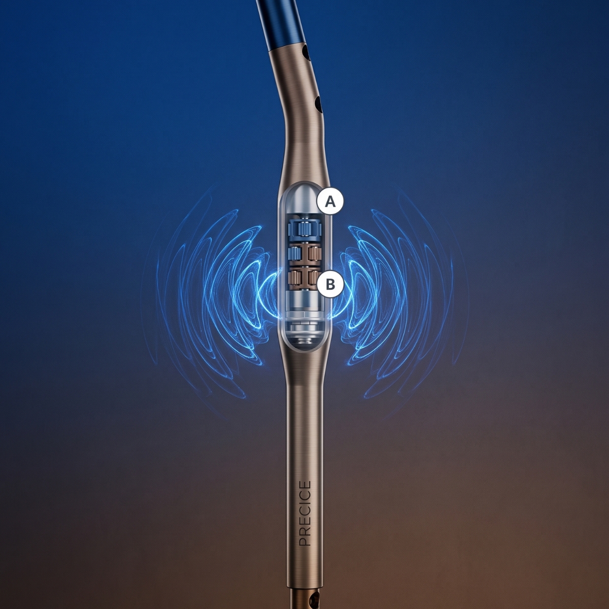
PRECICE – A Revolution in Limb Lengthening Without External Stabilizers