What are the most common causes of meniscus tears?
Pain in the shoulder or hip joint that worsens during daily activities may indicate a serious problem—damage to the labrum. The causes of this condition can vary. This small but extremely important structure ensures the stability and proper functioning of the joint. The labrum surrounds the acetabulum, deepening its shape and protecting the joint from instability, which is particularly important in the shoulder joint, where the head of the bone is significantly larger than the acetabulum. Labrum damage can lead to pain, limited mobility, and discomfort, especially in active individuals and athletes. Understanding the causes, symptoms, and methods of diagnosis and treatment of this condition is key to effective therapy and a return to full fitness.
From this article you will learn:
- Causes of meniscus tears – the most common risk factors
- Acute injuries: joint dislocations, landing on an outstretched hand, sudden twists and jerks
- Overuse injuries: repetitive overhead movements in athletes, intense physical exertion in runners and soccer players
- Congenital anatomical abnormalities: femoroacetabular impingement, hip dysplasia, labral hypertrophy
- Age-related natural wear and tear of the joint structure and joint instability
- Types of meniscus tears
- SLAP (Superior Labrum Anterior to Posterior) injuries of the shoulder joint – four classification types
- Bankart lesion affecting the anterior part of the glenoid cavity
- Damage to the hip joint labrum associated with femoroacetabular impingement
- Symptoms of a torn meniscus – how to recognize them
- Pain deep within the joint that is difficult to pinpoint, worsening with movements above shoulder level
- A sensation of popping, grinding, or locking in the joint
- Limited range of motion, weakened limb strength, and dead hand syndrome
- Pain in the groin area radiating to the knee joints in cases of hip injury
- Diagnosis of meniscal tears – a comprehensive approach
- Medical history and orthopedic examination, including clinical tests: O’Brien test, Speed test, Yergason test
- Imaging tests: shoulder ultrasound, contrast-enhanced MRI arthrography with approximately 80 percent accuracy
- The importance of magnetic resonance imaging in accurately visualizing damage to cartilage tissue
- Diagnostic challenges arising from the similarity of symptoms to those of other joint conditions
- Treatment methods for meniscus tears – effective strategies
- Conservative treatment: rest, limiting physical activity, cold compresses, pain relievers
- Physical therapy and rehabilitation involving exercises to strengthen and stabilize the joint
- Modern supportive therapies: PRP injections, shockwave therapy to stimulate tissue regeneration
- Surgical treatment: arthroscopy, suturing and fixation of the meniscus, resection of the damaged segment
- Summary
Causes of meniscus tears – the most common risk factors
Damage to the labrum can be caused by a variety of factors—ranging from acute mechanical trauma and chronic overuse to congenital anatomical abnormalities. Understanding these causes is crucial for both effective diagnosis and the implementation of appropriate treatment. It is worth noting that these injuries often coexist with other pathologies, such as changes within the rotator cuff, which further complicates the clinical picture.
Acute injuries: joint dislocations, landing on an outstretched hand, sudden twists and jerks
One of the most common causes of labral tears is acute trauma, which exerts a strong force on the joint over a short period of time. A shoulder dislocation—involving the displacement of the humeral head outside the glenoid cavity—poses a particularly serious threat to the integrity of the labrum. This type of injury most commonly occurs during contact sports such as rugby, judo, or basketball, as well as when the arm is raised excessively and forcefully above shoulder level.
Equally dangerous are falls onto an outstretched arm, during which the force of the impact is transmitted along the humerus directly to the joint. Sudden twists and jerks—characteristic, for example, of tennis, javelin throwing, or dynamic movements in basketball—generate shear forces that can tear the delicate structure of the labrum. In the case of the hip joint, a sudden twist of the hip during physical activity can similarly lead to acute damage to the acetabular labrum. Injuries of this type require rapid diagnosis and intervention to prevent further progression of the damage.
Overuse injuries: repetitive overhead movements in athletes, intense physical exertion in runners and soccer players
Not all labral tears result from a sudden, single-incident injury. Overuse microtrauma is another significant mechanism leading to labral tears—particularly in physically active individuals whose training involves repetitive, routine movements. Athletes who perform repeated overhead throws—such as baseball players, volleyball players, swimmers, or tennis players—expose the upper part of the shoulder joint labrum to systematic overuse, which over time leads to its degeneration and tear.
In the case of the hip joint, a similar risk applies to long-distance runners and soccer players, in whom repetitive rotational and flexion movements of the hip generate stresses that accumulate within the acetabular labrum. Microtraumas, though not individually noticeable, lead to a gradual weakening of the cartilaginous-fibrous structure, until even a minor exertion can cause complete damage. Therefore, regular monitoring of joint condition and proper recovery after exercise play a key role in preventing this type of injury.
Congenital anatomical abnormalities: femoroacetabular impingement, hip dysplasia, labral hypertrophy
Damage to the hip labrum is sometimes congenital or structural in nature—independent of the patient’s physical activity. Femoroacetabular impingement (FAI) is one of the most common anatomical causes of hip labral damage. It involves abnormal contact between the femoral head and the acetabulum, which leads to mechanical compression and gradual destruction of the labrum during hip movements. Two types of impingement are distinguished: the CAM type (abnormal shape of the femoral head) and the pincer type (excessive coverage of the head by the acetabulum), with both types coexisting in many patients.
Hip dysplasia—that is, abnormal formation of the acetabulum, resulting in insufficient coverage of the femoral head—is another significant risk factor. In such cases, the labrum bears excessive biomechanical loads, which accelerates its wear and tear and increases its susceptibility to injury. Labral hypertrophy—that is, its pathological enlargement—can, in turn, impair normal joint biomechanics and lead to chronic pain and limited mobility. All of these congenital anatomical abnormalities require individual assessment and often a specialized therapeutic approach.
Age-related natural wear and tear of the joint structure and joint instability
As we age, fibrocartilaginous tissues lose their elasticity and resistance to stress. Natural degenerative processes lead to the gradual wear and tear of the articular cartilage, making older adults particularly vulnerable to damage—even from relatively minor overuse or injuries. Reduced water content in the cartilage tissue, weakened collagen, and decreased cellular regenerative capacity cause the structure of the meniscus to become more brittle and prone to tears.
Joint instability—in both the shoulder and hip joints—is also a significant risk factor. When the muscles and ligaments surrounding the joint fail to provide adequate support, the head of the bone moves uncontrollably during movement, generating shear forces that act directly on the labrum. Shoulder joint instability—often a consequence of a previous dislocation—creates a vicious cycle in which labral damage exacerbates instability, and instability, in turn, contributes to further damage. Therefore, early diagnosis and treatment of joint instability are crucial for protecting the labrum from further damage.
Types of meniscus tears
A labral tear is not a homogeneous condition—in clinical practice, several distinct types are recognized, differing in location, mechanism of injury, and severity of the lesion. Knowledge of the specific types of injuries is crucial for the proper selection of treatment methods and rehabilitation planning. The most important of these are discussed below, with particular emphasis on SLAP lesions, Bankart lesions, and changes within the hip joint.
SLAP (Superior Labrum Anterior to Posterior) injuries of the shoulder joint – four classification types
SLAP (Superior Labrum Anterior to Posterior) tears are among the most commonly diagnosed types of labral tears in the shoulder joint. They affect the upper part of the glenoid labrum, where the tendon of the long head of the biceps brachii muscle attaches. These injuries are particularly common among athletes who perform repetitive overhead movements, such as pitchers, volleyball players, and swimmers, though they can also occur in people not involved in competitive sports.
In clinical classification, there are four basic types of SLAP lesions, which differ in the extent and nature of the anatomical changes:
- Type I – involves degeneration and fraying of the upper part of the labrum, while its attachment to the acetabulum remains intact. The attachment of the biceps tendon remains intact. This type of injury is most often associated with the natural aging process of the tissues and does not always require surgical intervention.
- Type II – involves the detachment of the upper portion of the labrum, along with the insertion of the long head of the biceps tendon, from the acetabulum. This is the most severe type of SLAP lesion and the one most often requiring surgical treatment. Surgical treatment involves suturing and reattaching the labrum to the acetabulum.
- Type III – characterized by a “basket-handle” tear of the labrum, in which part of the labrum remains attached to the acetabulum, while the detached fragment may move into the joint space, causing a sensation of locking or catching. The attachment of the biceps muscle is usually intact.
- Type IV is the most complex type of injury, in which the tear in the labrum extends to the tendon of the long head of the biceps brachii muscle. In this case, it may be necessary to remove the damaged portion of the tendon and reattach the labrum to the acetabulum.
It is worth noting that SLAP lesions may coexist with other conditions affecting the shoulder joint, particularly those involving the rotator cuff, which significantly complicates both diagnosis and treatment planning. These injuries represent complex orthopedic problems that require a personalized treatment approach.
Bankart lesion affecting the anterior part of the glenoid cavity
A Bankart lesion is another significant type of labral tear that affects the anterior and anteroinferior portions of the glenoid cavity. It is closely linked to the mechanism of shoulder dislocation—it most often occurs as a result of a sudden forward displacement of the humeral head, which causes the labrum to tear away from the edge of the glenoid cavity precisely at this location. A Bankart lesion is one of the main causes of recurrent shoulder instability, as it deprives the joint of a key stabilizing element.
In clinical practice, a distinction is made between classic Bankart lesions, which involve only the fibrocartilaginous structures of the labrum, and so-called bony Bankart lesions, in which a bony fragment of the acetabular rim is also damaged. The latter form is particularly severe and almost always requires surgical treatment. The mechanism underlying Bankart lesions therefore differs fundamentally from that of SLAP lesions—while SLAP lesions arise primarily from overuse and traction forces, Bankart lesions result from direct trauma and sudden displacement within the joint.
Damage to the hip joint labrum associated with femoroacetabular impingement
Labrum injuries are not limited to the shoulder joint—the hip labrum is equally susceptible to injury, although the mechanisms underlying these injuries are somewhat different. One of the most common causes of labral tears in the hip is femoroacetabular impingement (FAI), which involves abnormal contact between the femoral head and the acetabulum during lower limb movements. Abnormal joint geometry leads to mechanical irritation and gradual destruction of the labrum, particularly in its anterior and anterosuperior portions.
Injuries to the hip labrum can be either acute or chronic. In the case of an acute injury, the damage occurs suddenly, for example during a sudden twist of the torso or a fall. Chronic changes, on the other hand, develop gradually as a result of repeated microtraumas, which is characteristic of runners, soccer players, or dancers. Diagnosing these injuries can be difficult because the symptoms may resemble other conditions, such as snapping hip syndrome or adductor muscle injury, which requires detailed clinical and imaging examinations, including magnetic resonance imaging (MRI).
Symptoms of a torn meniscus – how to recognize them
A meniscus tear can manifest in many ways, and the nature of the symptoms depends primarily on the location of the injury—whether it affects the shoulder or hip joint. Symptoms can be nonspecific and difficult to definitively attribute to a specific structure, which often hinders prompt diagnosis. However, it is important to be aware of the warning signs that should prompt a consultation with a specialist. Untreated damage to the joint labrum can lead to accelerated degenerative processes and significant functional limitations.
Pain deep within the joint that is difficult to pinpoint, worsening with movements above shoulder level
One of the most characteristic symptoms of a torn shoulder labrum is pain felt deep within the joint, which patients often describe as difficult to pinpoint precisely. This is not superficial pain, but a sensation located deep within the joint structure, which can make it difficult for both the patient and the physician to identify during the initial clinical evaluation. The pain clearly worsens during movements above shoulder level—such as reaching for items on high shelves, throwing, or lifting objects overhead. Such symptoms may be chronic or worsen significantly after physical exertion, which is particularly burdensome for those who are active professionally and in sports. Symptoms may also accompany ordinary daily activities, such as combing hair or putting on a jacket, which significantly impacts the patient’s quality of life.
A sensation of popping, grinding, or locking in the joint
Another symptom to watch for is a characteristic sensation of catching, grinding, or a distinct locking sensation within the joint. These sensations result from a mechanical disruption of the joint’s normal kinematics, caused by the presence of a damaged or displaced piece of the labrum. Snapping and grinding in the shoulder joint can occur during both active and passive movements, and their intensity varies—sometimes they are accompanied by pain, while at other times they are merely an unpleasant but painless sensation. In the hip joint, similar symptoms may be confused with so-called snapping hip syndrome or adductor injury, which underscores the importance of a thorough differential diagnosis. A sensation of joint locking is particularly concerning, as it may indicate displacement of a labral fragment that mechanically restricts range of motion.
Limited range of motion, weakened limb strength, and dead hand syndrome
Damage to the labrum often leads to a gradual restriction of the joint’s full range of motion, which patients notice as an inability to perform certain movements or significant discomfort when attempting to reach the joint’s end-range positions. This is accompanied by weakness in the upper limb, particularly noticeable during abduction and external rotation of the arm. A particularly characteristic and concerning symptom is the so-called “dead arm syndrome,” which involves a sudden loss of control over the limb when the arm is in an extreme position. The patient then experiences temporary weakness, numbness, or helplessness in the limb, which is directly related to joint instability and a disruption of its normal biomechanics. This syndrome is particularly common in athletes who perform throws or serves, in whom the shoulder joint regularly reaches extreme ranges of motion. The functional limitations resulting from these symptoms can significantly impact the patient’s daily functioning and physical activity, making it difficult to perform both simple tasks and sports activities.
Pain in the groin area radiating to the knee joints in cases of hip injury
In the case of a hip labral tear, the clinical presentation is somewhat different and can be misleading for both the patient and the primary care physician. The main symptom is pain in the groin area, which patients often describe as deep, dull, or stabbing. The symptoms worsen during prolonged sitting, walking, or standing—that is, during activities that place stress on the hip joint for an extended period. Characteristically, the pain may radiate along the thigh all the way to the knee joints, which often directs diagnostic attention to other areas and delays the correct diagnosis. An additional symptom is stiffness in the hip joint, particularly noticeable after prolonged inactivity—such as when standing up from a chair or upon waking. Similar to the shoulder joint, a sensation of catching and limited range of motion may occur, confirming a mechanical dysfunction of the joint. Due to the similarity of these symptoms to other conditions, such as adductor injuries or degenerative changes, accurate diagnosis is essential for implementing appropriate treatment.
Diagnosis of meniscal tears – a comprehensive approach
Accurate diagnosis of a meniscal tear requires a multi-step diagnostic process that combines a detailed medical history, a physical examination, and modern imaging tests. Because the symptoms of a meniscus tear can resemble those of other joint conditions, precise diagnosis is crucial for implementing appropriate treatment and avoiding therapeutic errors. Each stage of the diagnostic process provides essential information that, when combined, allows for a complete and accurate determination of the nature and extent of the injury.
Medical history and orthopedic examination, including clinical tests: O’Brien test, Speed test, Yergason test
The first step in diagnosing a meniscus tear is a detailed medical history. The doctor asks the patient about the circumstances surrounding the onset of symptoms, their nature, location, and factors that exacerbate or alleviate the pain. Information regarding past injuries, physical activity, and any previous episodes of joint instability is important. Based on the medical history, it is possible to make a preliminary determination of the mechanism of injury and narrow down the range of suspected diagnoses.
An orthopedic examination includes an assessment of the joint’s range of motion, limb muscle strength, and the stability of joint structures. For the shoulder joint, standardized clinical tests are particularly useful, as they allow for the provocation of characteristic symptoms. The O’Brien test, also known as the active compression test, involves raising the limb to 90 degrees with the elbow flexed and the arm in internal rotation—the onset of deep pain within the joint suggests damage to the labrum or the long head of the biceps tendon. The Speed test evaluates the biceps tendon by having the patient lean on an outstretched upper limb while the forearm is in supination—pain in the anterior part of the shoulder indicates pathology within the labrum or tendon. The Yergason test, on the other hand, involves supinating the forearm with the elbow flexed while the examiner applies resistance—pain in the biceps groove indicates problems with the biceps tendon, often coexisting with SLAP-type labral tears. The results of these tests, when interpreted together, allow for a preliminary determination of the location and nature of the injury.
Imaging tests: shoulder ultrasound, contrast-enhanced MRI arthrography with approximately 80 percent accuracy
Imaging studies are an essential complement to the clinical examination and allow for direct visualization of damaged structures. Shoulder ultrasound is an accessible, non-invasive, and relatively quick procedure. It enables assessment of the rotator cuff, bursae, and the long head of the biceps tendon. However, due to its limited ability to penetrate deep into the joint, ultrasound does not allow for a complete assessment of the labrum—particularly its posterior and superior portions—and is therefore rarely sufficient as the sole imaging modality when labral injury is suspected.
A much more accurate tool is magnetic resonance arthrography (MRA), which involves performing an MRI scan after injecting contrast dye directly into the joint. The contrast agent fills the joint space, allowing even minor meniscal tears to be visualized—tears that might be invisible on a standard MRI scan. The accuracy of MR arthrography in diagnosing meniscal tears is approximately 80 percent, making it one of the most reliable tests available for diagnosing these conditions. The contrast agent highlights the boundaries between joint structures, enabling the physician to precisely assess the nature, location, and extent of the injury, which is essential for planning appropriate treatment.
The importance of magnetic resonance imaging in accurately visualizing damage to cartilage tissue
Magnetic resonance imaging (MRI) is the method of choice for diagnosing damage to cartilage tissues and the structure of the joint capsule. Unlike X-rays or computed tomography, MRI provides excellent imaging of soft tissues—cartilage, ligaments, tendons, and the joint capsule itself. Magnetic resonance imaging enables the detection of injuries that do not show up on X-rays, which is particularly important in the early stages of the condition, when therapeutic intervention can yield the best results.
Contrast-enhanced hip arthrography plays a key role in the diagnosis of hip labral tears, as it allows for the identification of pathological changes in the cartilage and labrum with high precision. MRI is also invaluable in assessing concomitant injuries—such as changes within the rotator cuff, articular cartilage damage, or pathologies of the long head of the biceps tendon—which may influence the choice of treatment. The MRI results, interpreted by an experienced radiologist in collaboration with an orthopedic surgeon, form the basis for making a treatment decision—whether conservative or surgical.
Diagnostic challenges arising from the similarity of symptoms to those of other joint conditions
Diagnosing a labral tear is a challenging process, as the symptoms of this condition can closely resemble those associated with other joint disorders. In the case of the hip joint, groin pain, a snapping sensation, or limited range of motion may suggest not only a labral tear but also snapping hip syndrome, adductor strain, bursitis, or degenerative joint changes. A similar situation applies to the shoulder joint, where symptoms of labral tear overlap with those of subacromial impingement syndrome, rotator cuff tendinitis, or joint instability.
The diagnostic challenge is compounded by the fact that meniscal tears may coexist with other conditions, meaning that isolating and identifying the specific sources of pain requires significant clinical experience. In such cases, imaging alone may not be sufficient—it must be correlated with the results of a physical examination and a detailed medical history. Sometimes, performing a diagnostic injection of an anesthetic into the joint proves helpful, as it confirms that the pain originates precisely from that area. A comprehensive, multi-step diagnostic approach—combining medical history, clinical tests, and imaging studies—is therefore essential for making an accurate diagnosis and implementing effective treatment for a meniscal tear.
Treatment methods for meniscus tears – effective strategies
The choice of the appropriate treatment method for a meniscus tear depends on many factors—the severity of the injury, its location, the patient’s age, their level of physical activity, and the time that has elapsed since the injury occurred. In most cases, treatment begins with conservative methods, and surgical intervention is considered only if there is no improvement or in cases of extensive damage. An individualized approach to each patient is crucial, as the causes and nature of meniscus injuries can vary significantly depending on the specific case.
Conservative treatment: rest, limiting physical activity, cold compresses, pain relievers
Conservative treatment is the first and often sufficient stage of therapy, especially in cases of partial labral tears. This approach is based on rest and limiting physical activity, which reduces stress on the joint and creates conditions conducive to the natural healing process of the tissues. In practice, this means temporarily avoiding activities that cause pain—especially movements above the shoulder line in the case of the shoulder joint or axial loads in the case of the hip joint.
Applying cold compresses in the first few days after an injury helps reduce swelling and inflammation within the joint. Compresses should be applied for 15–20 minutes several times a day, always with a protective layer between the ice and the skin to prevent frostbite. In the next stage of conservative treatment, pain relievers and anti-inflammatory medications are used, both orally and topically—their purpose is not only to relieve pain but also to reduce inflammation, which can hinder the healing process. If there is no improvement after three months of conservative treatment, doctors consider further therapeutic steps.
Physical therapy and rehabilitation involving exercises to strengthen and stabilize the joint
Physical therapy plays a key role in the treatment of labral tears, both as a standalone therapy and as an essential component of the rehabilitation process following surgery. The physical therapy program is always tailored to the specific type of injury, its location, and the patient’s current functional status. In the case of the shoulder joint, particular emphasis is placed on strengthening the rotator cuff muscles, which provide dynamic stabilization of the joint and relieve pressure on the damaged labrum.
Physical therapy involves a wide range of methods—from manual therapy, through proprioceptive and stabilization exercises, to joint mobilization techniques. Exercises that strengthen the muscles surrounding the joint are designed to compensate for the weakened passive stability that the labrum provided before the injury. Systematic rehabilitation not only helps reduce pain but also allows for the gradual restoration of full range of motion and strength in the limb. In cases of SLAP lesions and femoroacetabular impingement, physical therapy is an integral part of a comprehensive treatment plan.
Modern supportive therapies: PRP injections, shockwave therapy to stimulate tissue regeneration
Modern medicine offers an ever-wider range of options for supporting the body’s natural regenerative processes. PRP (platelet-rich plasma) injections are one of the most commonly used modern methods to support the treatment of damage to cartilage and soft tissues, including the joint capsule. PRP is obtained from the patient’s own blood through centrifugation and concentration of platelets, which contain growth factors that stimulate repair processes. Administering PRP directly to the area of the damaged structure accelerates healing, reduces inflammation, and can significantly improve joint function—especially in cases of partial meniscal tears.
Shockwave therapy is another modern method used in the rehabilitation of meniscal injuries, particularly effective in cases of chronic pain and overuse. This therapy works in multiple ways—it reduces tension in the muscles surrounding the joint, improves blood flow to the tissues, stimulates collagen production, and accelerates healing processes. Shockwave therapy is a non-invasive method, well-tolerated by patients, and can be used as a standalone supportive therapy or in combination with other conservative treatment methods. The combination of PRP injections with shockwave therapy and physical therapy creates a comprehensive treatment approach that, in many cases, makes surgery unnecessary.
Surgical treatment: arthroscopy, suturing and fixation of the meniscus, resection of the damaged segment
In cases where conservative treatment does not produce the desired results or when the damage to the joint capsule is extensive, surgical intervention becomes necessary. The preferred surgical method is arthroscopy—a minimally invasive technique that involves inserting a miniature camera and surgical instruments into the joint through small incisions. Arthroscopy minimizes trauma to the tissues surrounding the joint, shortens the duration of hospitalization and recovery, and reduces the risk of complications compared to traditional open surgery.
Depending on the nature and extent of the injury, various procedures are performed during arthroscopy. Suturing and reattaching the labrum to the rim of the acetabulum is recommended for injuries in which the labrum’s structure is sufficiently intact—a typical example is the treatment of type II SLAP lesions and Bankart lesions. For this purpose, implants made of biomaterials are increasingly used instead of metal anchors, which reduces the risk of degenerative changes in the joint in the long term. In contrast, resection of the damaged portion of the labrum is performed in situations where the tissue is too damaged to be repaired—this primarily applies to certain types of SLAP lesions and chronic injuries accompanied by degenerative changes. The decision regarding the choice of a specific surgical procedure always takes into account the patient’s age, physical activity level, number of previous dislocations, and the overall condition of the joint.
Summary
A meniscus tear is a condition that can result from acute trauma, chronic microtrauma, or congenital anatomical abnormalities. Characteristic symptoms, such as pain, joint catching, or limited range of motion, require precise diagnosis, often involving imaging tests. Treatment may be conservative or surgical, but properly managed rehabilitation plays a key role in enabling a full return to function. These symptoms should not be ignored, as untreated labral damage can lead to serious problems, including accelerated progression of osteoarthritis. Early diagnosis and a comprehensive approach to treatment significantly increase the chances of a full recovery. If you experience pain in your shoulder or hip joint, a sensation of catching, or limited mobility, don’t delay—consult an orthopedist or physical therapist. In this regard, paleyeurope.com is an excellent resource, offering professional care and effective rehabilitation methods that will help you return to full activity and enjoy good health every day.
See other entries
What is SEMLS surgery, and who needs it?
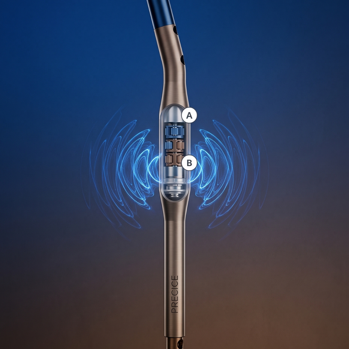
PRECICE – A Revolution in Limb Lengthening Without External Stabilizers